Stress

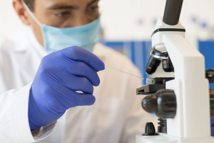
Close-up of scientist hands with microscope

A recent stem cell study has shown that SARS-CoV-2, the novel coronavirus, can infect heart cells via the same receptor present in the lungs. This may be responsible for the cardiac complications associated with COVID-19.

Experts initially thought that COVID-19 was a respiratory disease, with symptoms including cough, shortness of breath, and pneumonia. However, more recent evidence into COVID-19 shows that the disease can also cause neurological and cardiac symptoms.

Physicians have reported changes to the circulatory system in people with COVID-19, sometimes leading to blood clots, as well as cardiac complications, such as changes to the heart rhythm, damage to heart tissue, and heart attacks.

Although there is widespread agreement that COVID-19 is a risk to the heart, whether these symptoms are due to the virus directly or a consequence of other disease processes, such as inflammation, has been unclear.

In a new study appearing in the journal Cell Reports Medicine, scientists have helped resolve this mystery by showing that SARS-CoV-2 can infect heart cells and change their function.

Their findings, from experiments in human stem cells, suggest that the cardiac symptoms of COVID-19 may be the direct result of the infection of heart tissue.

SARS-CoV-2 behaviour in heart cells

The scientists used a type of stem cell called induced pluripotent stem cells (iPSCs) to generate heart cells.

Scientists can create iPSCs from a person’s skin cells and then reprogram them to become any cell type in the body. They provide a useful tool for research into human disease and a way to test new treatments.

In this study, the team programmed the iPSCs to become heart cells and later incubated them with SARS-CoV-2. Using microscopes and genetic sequencing techniques, the researchers found that SARS-CoV-2 could directly infect the heart cells.

They also showed that the virus can rapidly divide inside heart cells, which caused changes to the heart’s ability to beat after a period of under 3 days.

“We not only uncovered that these stem cell-derived heart cells are susceptible to infection by [the] novel coronavirus, but that the virus can also quickly divide within the heart muscle cells,” explains first study author Dr. Arun Sharma, a research fellow at the Regenerative Medicine Institute of Cedars-Sinai Medical Center in Los Angeles, CA.

The role of the ACE2 receptor

Additional experiments focused on the different genes expressed by heart cells before and after the virus infected them. These studies showed activation of the innate immune response and antiviral clearance pathways to help fight the virus.

However, how does the virus get into the heart in the first place? The researchers suggest that one way in which it gains access may be by using angiotensin-converting enzyme 2 (ACE2). This is the same receptor the virus uses to infect cells in the lungs.

Importantly, studies have shown that treatment with an ACE2 antibody can help stop SARS-CoV-2 from replicating and save cells in the heart.

“By blocking the ACE2 protein with an antibody, the virus is not as easily able to bind to the ACE2 protein, and thus cannot easily enter the cell. This not only helps us understand the mechanisms of how this virus functions, but also suggests therapeutic approaches that could be used as a potential treatment for SARS-CoV-2 infection.” – Dr. Arun Sharma

The importance of these findings

The researchers suggest that scientists could use stem cell-derived heart cells to screen new drugs and find compounds able to stop the infection of heart cells.

“This key experimental system could be useful to understand the differences in disease processes of related coronaviral pathogens SARS and MERS,” adds study author Dr. Vaithilingaraja Arumugaswami, an associate professor at the University of California, Los Angeles.

There are some limitations to this approach, however. These include the fact that stem cell-derived heart cells are not exactly the same as the real thing.

The researchers also studied the cells in a dish, an isolated system lacking the immune interactions that would occur in the human body.

Nevertheless, the experiments clearly showed that the cells became infected with SARS-CoV-2, which is in line with some clinical data showing the virus in the hearts of people who died from COVID-19.

Executive Director, ISN Medical laboratory Mr. Felix Ofungwu, has reiterated need to train and retrain medical laboratory scientists in the country as it remains the best way to reduce misdiagnoses of diseases.

He however, explained that causes of misdiagnosis include delay during the diagnosis process, human errors, quackery in the profession and personal biases about patients among others are common causes of misdiagnosis, which are increasing in the country.

He stated this during a press briefing to announce ISN Medical Laboratory Scientist of the Year award. He said the ward is a way of recognising and rewarding the efforts of medical Laboratory Scientist across the country including front line health workers in the ongoing fight against coronavirus, held at Ilupeju, Lagos.

He noted that the ISN award would run from June to November 2020 and entry is open to practicing licensed medical laboratory scientists while an erudite panel of judges will execute grading and selection of the winner in September while the grand final and presentation of prizes would hold in November.

The immediate past President of the Medical Laboratory Science Council of Nigeria (MLSCN), Dr. Raheed Toyosi, said MLSCN is in full support of the project adding that the eligible, participant must have the requisite academic qualifications, must be a registered member of the Medical Laboratory Science Council of Nigeria (MLSCN), and must be working in a registered medical laboratory or hospital. This is as much a recognition of the laboratory in, which the scientist works as it is about the science.

He noted that overall winner of the award will receive a grand prize of one million naira with a capacity development training with international partners and a donation of medical lab equipment/reagents worth one million naira to the lab or hospital where he or she works.

Olusola Malomo, a leading Lagos nutritionist/Publicity Secretary of Nutrition Society of Nigeria, has called for regular intake of 100% fruit juice and regular exercise among others, as measures to boost the immune system and combat the Coronavirus Disease (COVID-19) pandemic. He gave the advice at his monthly healthy living dialogue.

While noting that COVID-19 is a respiratory infection which causes fever, tiredness, sore throat and in severe cases, shortness of breath and respiratory difficulty, he said that the World Health Organization (WHO) and other global public institutions have confirmed that the severity of the COVID-19 infection is linked with the overall state of the body’s immune system. He stated that this is the reason why people with pre-existing health challenges are most vulnerable to the contagion.

Quoting the Harvard Medical School in its Harvard Health Publishing, Malomo revealed that deficiency in Zinc, Iron, Copper, Folic Acid, Vitamins A, B6, C (which is contained in large quantity in fruit juice) and E have negative impacts on immune responses.

He said: “CNN reported that the United States retail sales of orange juice jumped about 38 percent in the four weeks ending on March 28, 2020, when compared to the same period last year. Also, the Florida Department of Citrus disclosed that there was a spike in demand for 100% orange juice within the same period.”

“During the five weeks the Federal Capital Territory, Lagos and Ogun were on lockdown, shops and supermarkets had run out of orange fruit juice stock in the first two or three weeks of the restriction.”

“In fact, less than a week into the lockdown, my favorite Chivita 100% Real Orange Fruit Juice which would have made the stay-at-home less boring for me, had rapidly been purchased off the shelves in Lagos. The scramble for unavailable raw fruits in the open markets started in earnest as the restriction on movement cut off a large chunk of the supplies.”

The expert, however, stated that fruit juice intake is just one of the many ways to boost one’s immune system. Others measures according to him include getting adequate sleep, thorough cooking of meat, maintaining good hygiene, moderate intake of alcohol and exercising regularly.

“Regular exercise is an essential component of healthy living. It improves cardiovascular function, helps control body weight, removes toxins in the body, increases blood circulation and lowers blood pressure. The result of these is a strengthened immune system that protects the body against infections and diseases. Regular exercise is most valuable when one shuns junk food and sticks to healthy eating habits and a balanced diet,” he noted. It is difficult to ascertain the level of good hygiene maintained by those who sell raw fruits, and considering that good hygiene is essential to combat COVID-19, Malomo said pure fruit juices, where available, can be substituted for raw fruits. He, however, cautioned against the consumption of brands whose production processes are not trusted.

A new research published last week in the Journal of Physiology showed that 12 weeks of easy-to-administer passive stretching helps improve blood flow by making it easier for your arteries to dilate and decreasing their stiffness.

Passive stretching differs from active stretching in that the former involves an external force (another person or gravity) stretching you, whereas active stretching is performed on your own. The changes they observed in blood vessels could have implications for diseases, including the number one global killer, heart disease.

Researchers at the University of Milan assigned 39 healthy participants of both sexes to two groups. The control group didn’t undergo any stretching. The experimental group performed leg stretches five times a week for 12 weeks. Researchers evaluated the effect of passive stretching on the blood flow locally and in the upper arm. They found that the arteries in both the lower leg and upper arm had increased blood flow and dilation when stimulated, along with decreased stiffness.

Both of these changes may have implications for diseases such as heart disease, stroke and diabetes as they are characterized by changes in blood flow control, due to an impaired vascular system.

If this study is replicated in patients with vascular disease, it could indicate whether or not this training method could serve as a new drug-free treatment for improving vascular health and reducing disease risk, especially in people with lower mobility.

Moreover, stretching may also be used during hospitalisation or after surgical interventions, to preserve the vascular health when patients have low mobility. Carers or family members can also perform it at home.

An author on the paper, Emiliano Ce, said this new application of stretching is especially relevant in the current pandemic period of increased confinement to homes, where the possibility of performing beneficial training to improve and prevent heart disease, stroke and other conditions is limited.

Also, another research suggests that exercise can slow or prevent the development of macular degeneration and may benefit other common causes of vision loss, such as glaucoma and diabetic retinopathy.

The new study from the University of Virginia School of Medicine found that exercise reduced the harmful overgrowth of blood vessels in the eyes of lab mice by up to 45 per cent. This tangle of blood vessels is a key contributor to macular degeneration and several other eye diseases.

The study represents the first experimental evidence showing that exercise can reduce the severity of macular degeneration, a leading cause of vision loss, the scientists report. Ten million Americans are estimated to have the condition.

Researcher at the UVA’s Center for Advanced Vision Science, Dr. Bradley Gelfand, said: “There has long been a question about whether maintaining a healthy lifestyle can delay or prevent the development of macular degeneration. The way that question has historically been answered has been by taking surveys of people, asking them what they are eating and how much exercise they are performing.

“That is basically the most sophisticated study that has been done. The problem with that is that people are notoriously bad self-reporters … and that can lead to conclusions that may or not be true. This [study] offers hard evidence from the lab for very first time.”

Enticingly, the research found that the bar for receiving the benefits from exercise was relatively low — more exercise didn’t mean more benefit. “Mice are kind of like people in that they will do a spectrum of exercise. As long as they had a wheel and ran on it, there was a benefit,” Gelfand said. “The benefit that they obtained is saturated at low levels of exercise.”

An initial test comparing mice that voluntarily exercised versus those that did not found that exercise reduced the blood vessel overgrowth by 45 per cent. A second test, to confirm the findings, found a reduction of 32 per cent.

The scientists aren’t certain exactly how exercise is preventing the blood vessel overgrowth. There could be a variety of factors at play, they say, including increased blood flow to the eyes.

Gelfand, of UVA’s Department of Ophthalmology and Department of Biomedical Engineering, noted that the onset of vision loss is often associated with a decrease in exercise. “It is fairly well known that as people’s eyes and vision deteriorate, their tendency to engage in physical activity also goes down,” he said. “It can be a challenging thing to study in older people. … How much of that is one causing the other?”

The researchers already have submitted grant proposals in hopes of obtaining funding to pursue their findings further.

“The next step is to look at how and why this happens, and to see if we can develop a pill or method that will give you the benefits of exercise without having to exercise,” Gelfand said. “We’re talking about a fairly elderly population [of people with macular degeneration], many of whom may not be capable of conducting the type of exercise regimen that may be required to see some kind of benefit.” (He urged people to consult their doctors before beginning any aggressive exercise program.)

Gelfand, a self-described couch potato, disclosed a secret motivation for the research: “One reason I wanted to do this study was sort of selfish. I was hoping to find some reason not to exercise,” he joked. “It turned out exercise really is good for you.”

Photo series of a virtual dinner date with video call during lockdown.

New research suggests that low-to-moderate alcohol consumption may reduce cognitive decline in older adults.

A new study has found that low-to-moderate alcohol consumption may slow cognitive decline in adults of middle age or older.

The research, which appears in the journal JAMA Open Network, lays the ground for future research to corroborate these findings and determine the underlying mechanism for the relationship.

Alcohol and health

Drinking alcohol is a central part of many cultures across the world. Beyond the immediate adverse effects of a hangover, drinking alcohol in excess has associations with various poor health outcomes.

According to the Centers for Disease Control and Prevention (CDC), excessive alcohol use includes heavy drinking (more than eight or 15 drinks a week for women and men, respectively), binge drinking (drinking four or five drinks in about 2 hours for women and men, respectively), or drinking while underage or pregnant.

The CDC define a drink as 14 grams of pure alcohol. This amount roughly equates to a 12-ounce (oz) can of 5% beer, a 5-oz glass of 12% wine, a 4-oz glass of 15% wine, or a single 1.5-oz shot of 40% distilled spirit or liquor.

The CDC note that excessive alcohol drinking is associated with various health problems. These include:

  • damage to the liver
  • inflammation of the pancreas
  • increased risk of cancer
  • hypertension
  • psychological disorders
  • unintentional injuries that a person sustained while drunk
  • harm to the fetus if a woman drinks while pregnant
  • sudden infant death syndrome
  • alcohol use disorder

Research has shown that the misuse of alcohol is a leading cause of illness and death and that any level of alcohol drinking, particularly in men who drink spirits, increases the risk of high blood pressure and stroke.

However, there is also evidence that low-to-moderate alcohol consumption — fewer than eight or 15 standard drinks in the United States per week for women and men, respectively — can have a beneficial effect on health, including various cardiovascular diseases.

The effects of low-to-moderate alcohol consumption on cognitive decline in later life have been mixed. Some research has found evidence that it may be protective, while other research suggests that it is probably not harmful. Conversely, some studies have found that it can exacerbate poor brain health.

The authors of the present study noted that previous studies exploring the relationship between alcohol consumption and cognitive decline are often limited in two ways.

First, they typically rely on single measures of cognitive decline, when it can present in a variety of ways. Second, they typically take into account net follow-up time to cognitive decline, despite the decline often being greater or lesser depending on age rather than general time passed.

As a consequence, the authors of the present research developed their study to take these factors into account, with the aim of better understanding the effects of low-to-moderate alcohol consumption on cognitive decline in older age.

About 20,000 participants

The authors drew on data from the Health and Retirement Study, which was a longitudinal study involving about 20,000 adults of middle age or older living in the U.S.

The sample that the researchers used was representative of the U.S. population, and they tracked the participants for an average time of 9.1 years.

The team regularly assessed the participants’ cognitive functioning during this time, focusing on three measures: total word recall, mental status, and vocabulary. They split the participants into those with persistently high scores across time and those with persistently low scores across time.

The participants also gave information on how many alcoholic drinks they consumed on average. During the data analysis, the researchers separated them into three groups: those who never drank, those who used to drink, and those who currently drink.

They then further classified drinkers as low-to-moderate drinkers (fewer than eight or 15 drinks per week for women and men, respectively), or heavy drinkers (more than moderate drinking).

Low-to moderate drinking may protect against cognitive decline

After analyzing the data, the researchers found that low-to-moderate drinking was associated with better cognitive outcomes across all three measures for middle-aged and older adults.

As other factors could be responsible for this association, the researchers took age, sex, race/ethnicity, educational level, marital status, and body mass index (BMI) into account in the analysis. The association between consistently high cognitive scores and low-to-moderate drinking persisted.

While this was true in general across the study, the researchers found that the association between low-to-moderate alcohol consumption and protection against cognitive decline was stronger for white participants than for Black participants. However, there were fewer Black participants, and this could account for the lack of strong association.

The researchers also noted that about 5% of women and 15% of men drank excessive amounts of alcohol, meaning that public health awareness campaigns that focus on this issue are still crucial. Drinking more than 14 drinks (self-defined) had worse cognitive outcomes for vocabulary, while more than 10 drinks impacted negatively on word recall.

Future research

While the study demonstrates an association between low-to-moderate alcohol drinking and protection against cognitive decline in later life, it is not yet clear why this is the case. According to study author Dr. Ruiyuan Zhang of the Department of Epidemiology and Biostatistics, University of Georgia College of Public Health, Athens, and his co-authors:

“The role of alcohol drinking in cognitive function may be a balance of its beneficial and harmful effects on the cardiovascular system. Among low-to-moderate drinkers, the beneficial effects may outweigh the harmful effects on the cardiovascular system.”

Future research to corroborate the study’s findings is important, given the previous conflicting research on the topic. In addition, if research conclusively demonstrates low-to-moderate alcohol consumption as protective against cognitive decline in older age, future research to help understand why this is the case is important so that clinicians can best advise their patients.

There is “strong evidence” that COVID-19- positive mothers can pass the virus on to their unborn infants, scientists said Thursday, in findings that could affect how pregnant women are shielded during the pandemic.

While there have been isolated cases of babies infected with the virus, the findings show the strongest link yet between mother and infant transmission.

Researchers in Italy studied 31 pregnant women hospitalised with COVID-19 and found the virus in an at-term placenta, umbilical cord, the vagina of one woman and in breast milk.

They also identified specific COVID-19 antibodies in the umbilical cords of several pregnant women as well as in milk specimens.

Claudio Fenizia, from the University of Milan and lead study author, said the findings “strongly suggest” that in-vitro transmission is possible.

“Given the number of infected people worldwide, the number of women that could be affected by this could be potentially very high,” he told AFP.

Fenizia stressed that none of the infants born during the study period tested positive for COVID-19.

“Although in utero transmission seems to be possible, it is too early to clearly assess the risk and potential consequences,” he said.

The World Health Organization said last month that new mothers infected with COVID-19 should continue breastfeeding.

“We know that children are at relatively low-risk of COVID-19, but are at high risk of numerous other diseases and conditions that breastfeeding prevents,” said WHO chief Tedros Adhanom Ghebreyesus.

Among other findings, the team identified a specific inflammatory response triggered by COVID-19 in the women’s placenta and umbilical cord blood plasma.

Fenizia said that the women studied were all in their third trimester, given the timeframe of Italy’s epidemic, adding that more research is currently underway among COVID-19-positive women in the early stages of pregnancy.

“Our study is aimed at raising awareness and inviting the scientific community to consider the pregnancy in positive women as an urgent topic to further characterise and dissect,” he said.

“I believe that promoting prevention is the safer advice we could possibly give right now for these patients.”

The study was released during a week-long International AIDS Conference, held online for the first time in its history due to the pandemic.

Scientists have discovered a way to track changes in sound processing in the brains of people with HIV. Their test provides a simple technique for studying how HIV affects the central nervous system.

Due to advances in antiretroviral drug treatment over the past 20 years, most people with HIV can now expect to live long, healthy lives.

However, even if treatment successfully brings the virus under control, people can experience cognitive issues as a result of HIV causing damage to their central nervous system.

Up to 45% of people with HIV may develop these difficulties, which are known collectively as HIV-associated neurocognitive disorder (HAND).

Although the cause of HAND remains unknown, scientists have several theories. They speculate, for example, that it may be the result of chronic inflammation, lingering damage from the initial infection, or a toxic effect of antiretroviral drugs.

Alternatively, low levels of the virus may persist in the brain as a result of the blood-brain barrier limiting the passage of antiretroviral drugs into the brain.

The signs and symptoms of HAND can be subtle, which makes it challenging for doctors to diagnose early and monitor.

Understanding speech

One of the problems that people with HIV sometimes report is difficulty understanding speech in the presence of background noise.

Researchers at the Geisel School of Medicine at Dartmouth in Hanover, NH, led a team that studied the hearing of a group of people with HIV in Dar es Salaam in Tanzania.

They collaborated with scientists from the Muhimbili University of Health and Allied Sciences in Dar es Salaam and neuroscientists from Northwestern University in Evanston, IL.

“Initially, we thought we’d find that HIV affects the ear, but what seems to be affected is the brain’s ability to process sound,” says study co-leader Dr. Jay Buckey Jr., a professor of medicine at Geisel.

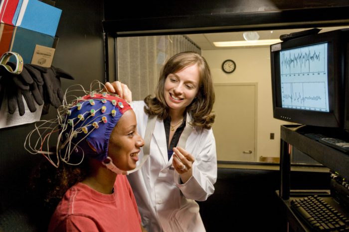
To test this idea, they used scalp electrodes to monitor the brain waves of 68 people with HIV and 59 people without as they listened to sounds.

Specifically, they recorded a brain response known as the speech-evoked frequency-following response (FFR).

Certain features of brain waves, such as their timing and amplitude, reveal how faithfully the brain encodes an auditory stimulus. The FFR has proven particularly useful for investigating how the brain processes information in highly complex sounds, such as music and speech.

The precision and accuracy of the FFR can improve as a result of experience, such as in trained musicians, or worsen as a result of illness or damage, such as concussion.

“There are many acoustic ingredients in speech, such as pitch, timing, harmonics, and phrase,” says study co-leader Nina Kraus, Ph.D., a professor of communication sciences and neurobiology at Northwestern.

“The FFR enables us to play speech sounds into the ear of study participants and figure out how good a job the brain is doing processing these different acoustic ingredients.”

Common speech sounds

The researchers recorded the FFR while the study participants listened to common speech sounds, such as “ba,” “da,” and “ga.”

They looked at how well the signal encoded two acoustic features of speech, known as the harmonics corresponding to the first formant and fundamental frequency.

The harmonics corresponding to the first formant identify the units of sound, or “phonemes,” that differentiate one word from another, whereas the fundamental frequency reflects the frequency at which the vocal cords vibrate, which helps identify the speaker.

The brains of people with HIV had similar responses to the brains of people without HIV at processing the fundamental frequency.

They also performed well on standard tests of hearing, but the FFR revealed that the brains of people with HIV were worse at encoding the first formant.

In other words, they had normal hearing thresholds, but their brains could not distinguish between the sounds of different words as accurately.

“When the brain processes sound, it’s not like a volume knob where all of the acoustic ingredients are either processed well or poorly,” Kraus explains. “With the FFR, we’re able to see which aspects of auditory processing are affected or diminished and ask, ‘[I]s there a specific neural signature that aligns itself with HIV?’”

The researchers have published their study in the journal Clinical Neurophysiology.

Early aging

The researchers note that as people get older, even if their hearing remains normal, their brains get worse at processing the same speech features that the brains of people with HIV seem to struggle with.

They write that HIV-associated cognitive problems may therefore be akin to early aging.

In the future, they hope that other scientists will use the FFR not only to study brain dysfunction associated with HIV, but also other conditions, such as concussion and Alzheimer’s disease.

Standard tests of cognitive function involve giving people tasks, such as solving math problems or remembering a list of words. However, these are dependent on the language people speak and their culture.

“What’s significant about our results is that the test doesn’t require any actions on the [subject’s] part. It’s recorded passively — subjects can even sleep or watch a movie,” says Dr. Buckey. “We think the FFR holds a lot of promise as a way to assess the brain easily and objectively.”

In their paper, the researchers note that the test could prove particularly useful in parts of the world with the most HIV infections and limited healthcare resources.

They write, “While previous neuroimaging and electrophysiological studies have shown differences between HIV+ and HIV- individuals, these approaches are difficult to transport to resource-limited settings such as sub-Saharan Africa, where >70% of the world’s HIV+ population lives and where there is some evidence that neurocognitive symptoms are more severe.”

“Thus, the FFR holds promise as a research tool to further study [central nervous system] health, particularly in resource-limited settings.”

The study authors acknowledge that research projects involving more participants will be necessary to confirm their results. They say that it would also be interesting to track changes in the FFR from the early stages of infection onward.

Finally, in light of evidence suggesting that different strains of the virus may have differing neurological effects, they would like to see studies that compare FFR tests in different populations around the world.

by -
0 533
Close-up of woman's hand holding Vitamin D supplements

A review from health experts in the United Kingdom has found no evidence that vitamin D deficiency is an independent risk factor for COVID-19. However, the country’s National Health Service recommend taking a daily vitamin D supplement to compensate for any lack of sunlight exposure during lockdown.

Vitamin D is vital for maintaining healthy bones and muscles. There is also some evidence that it may help protect against viral respiratory infections and play a regulatory role in the body’s immune response.

This has led to a proposal that taking vitamin D supplements could help prevent or even treat COVID-19, the primarily respiratory disease caused by the virus SARS-CoV-2.

While no clinical trials have tested the vitamin’s efficacy as a treatment or a preventive measure, several studies have found an association between low levels of vitamin D and COVID-19.

However, a review of five of these studies by the U.K.’s National Institute for Health and Care Excellence (NICE), which compiles guidelines on best practices, concludes that the studies provide no evidence that vitamin D levels influence the risk of getting COVID-19 or dying as a result.

None of the five studies had been designed to investigate the effects of the supplements on the risk of the infection or as a treatment for the disease, the authors point out. Either investigation would have required an intervention study, such as a randomized controlled trial.

The existing studies, therefore, provide no insights into the vitamin’s efficacy, appropriate doses, or possible adverse effects as a means of treating or preventing COVID-19.

Confounding factors

Moreover, only one study in the review accounted for confounding factors, which may provide alternative explanations for the observed associations between vitamin D status and COVID-19.

Higher body mass index (BMI), older age, and socioeconomic deprivation, for example, are all factors that could affect both the risk of COVID-19 and levels of the vitamin.

This makes it impossible to draw any firm conclusion about whether having a lack of the vitamin increases the risk of contracting the virus that causes COVID-19 or dying as a result of the disease.

One of the studies, reported by Medical News Today, found an association between average levels of vitamin D and numbers of COVID-19 cases and deaths by country. But the research had limitations — it did not, for example, account for the proportions of older people in these populations.

The only study reviewed by NICE that had adjusted for potential confounding factors — including preexisting illnesses, sociodemographic factors, ethnicity, and BMI — found no independent association between COVID-19 and vitamin D levels.

As the authors of the review observe:

“There is no evidence to support taking vitamin D supplements to specifically prevent or treat COVID-19. However, all people should continue to follow U.K. government advice on daily vitamin D supplementation to maintain bone and muscle health during the COVID-19 pandemic.”

The review focused exclusively on studies that had been peer reviewed and published in scientific journals.

“Vitamin D enthusiasts will point to a large number of other studies, published on preprint websites, that were not included in this review,” says Prof. Adrian Martineau, a clinical professor of respiratory infection and immunity at Queen Mary University of London, who was not involved in the NICE review.

“However, these studies have not yet undergone peer review, so their findings should not be relied on to guide clinical practice or public health policy.”

Sunlight exposure

Only about 10% of the vitamin D in our bodies originates from food. The remainder is synthesized in the skin through exposure to ultraviolet light in sunlight.

Research suggests that the U.K. population has one of the highest levels of vitamin D deficiency in Europe.

The country’s National Health Service, therefore, recommend that people take a daily supplement containing 10 micrograms of vitamin D during the autumn and winter months, when sunlight exposure may be limited.

But its latest advice says that people should consider taking a supplement during the summer months, as well, if they are spending more of their time indoors as a result of the pandemic.

The aim of this recommendation is to support bone and muscle health, rather than to protect against COVID-19.

Call for research

In a preprint review, The Royal Society, in London, has called for more research into the possibility that vitamin D deficiency predisposes people to COVID-19.

The review cites several lines of evidence to suggest that people with the deficiency may be more susceptible to the disease:

“Vitamin D deficiency is associated with an increased risk of both respiratory viral infections and inflammatory conditions. Vitamin D has an important regulatory role in the human immune system, so a deficiency of vitamin D is likely to cause immune dysregulation, which may reduce the first line of our defense against COVID-19. It is therefore biologically plausible that vitamin D deficiency may contribute to susceptibility to COVID-19 infection. However, there is no direct causal link yet between vitamin D deficiency and increased susceptibility to COVID-19.”

The authors point out that vitamin D deficiency is more common among older people, those who are Black or of Asian origin, and those who have obesity. These groups of people are also known to have an increased risk of developing severe COVID-19.

Nonetheless, the scientists emphasize that “Correlations are not the same as causality.”

A laboratory study has found that the asthma drug salbutamol prevents the formation of tangles of fibrous protein that are a hallmark of Alzheimer’s disease. The next step will be to test the drug in animal models of the disease.

About 50 million people worldwide have dementia, and every year, there are nearly 10 million new cases, according to the World Health Organization (WHO).

They note that the most common form of dementia is Alzheimer’s disease, which accounts for 60–70% of all cases.

In the United States, the National Institute on Aging estimate that more than 5.5 million people have Alzheimer’s disease. Most of them are over 65 years of age.

The disease is a neurological disorder in which the death of brain cells results in progressive memory loss and cognitive decline.

While existing drug treatments help reduce the symptoms of Alzheimer’s disease and improve people’s quality of life, they neither slow its progression nor cure it.

The brains of people with the condition contain distinctive plaques between nerve cells, as well as clumps of fibers known as neurofibrillary tangles inside the cells.

The plaques consist of a protein called beta-amyloid, while another protein called tau makes up the tangles.

Promising drug target

After clinical trials found that drugs that clear beta-amyloid from the brain failed to slow the disease’s progression, attempts to find a treatment have shifted to tau.

Researchers at Lancaster University in the United Kingdom believe that tau could be a more promising drug target for Alzheimer’s disease. They point to previous research, which found that in the absence of the tau neurofibrillary tangles, beta-amyloid does not seem to harm nerve cells.

Also, the number of tangles in the brain appears to be a much better indicator of the severity of the disease than the number of amyloid plaques.

In healthy brain cells, tau proteins help stabilize the internal network of microscopic tubes, or “microtubules,” that transports nutrients and other molecules around nerve cells.

In Alzheimer’s disease, these tau molecules break away from the microtubules and begin to stick together to form threads and, eventually, tangles. In turn, these disrupt the microtubule transport network.

The scientists from Lancaster University believe that compounds that prevent tau molecules from aggregating in this way could make promising treatments for Alzheimer’s disease.

Giant microscope

To screen more than 80 compounds for their ability to block the formation of tangles, the researchers used a powerful technique — synchrotron radiation circular dichroism — for imaging structural changes in proteins.

This technique involves illuminating samples with beams of light 10 billion times brighter than the sun. The U.K.’s Diamond Light Source (DLS) in Oxfordshire, which experts have likened to a giant microscope, generated this light for the study.

One of the compounds that the DLS identified was the hormone epinephrine, which stabilized tau proteins and prevented them from forming tangles.

The body rapidly metabolizes epinephrine, however, so the researchers went on to screen four existing drugs with very similar chemical structures.

Of these, two were effective: a drug called dobutamine, which doctors use to treat heart attacks and heart failure, and salbutamol (also known as albuterol), which is available under the brand name Ventolin for treating asthma.

The scientists ruled out dobutamine as a practical treatment for Alzheimer’s disease because it requires injection, and its effects are very short-lived.

Further tests on salbutamol suggested that it binds to individual tau molecules, preventing them from forming “nuclei” around which other protein molecules can aggregate.

The researchers published their findings in the journal ACS Chemical Neuroscience.

Early days

“This work is in the very early stages, and we are some way from knowing whether or not salbutamol will be effective at treating Alzheimer’s disease in human patients,” says Prof. David Middleton, one of the authors.

“However, our results justify further testing of salbutamol and similar drugs in animal models of the disease and, eventually, if successful, in clinical trials.”

Salbutamol is on the WHO’s Model Lists of Essential Medicines and is the 10th most commonly prescribed medication in the U.S.

Dr. David Townsend, who led the research, says that it demonstrated the potential benefits of finding new applications for existing drugs with proven safety records:

“Salbutamol has already undergone extensive human safety reviews, and if follow-up research reveals an ability to impede Alzheimer’s disease progression in cellular and animal models, this drug could offer a step forward, whilst drastically reducing the cost and time associated with typical drug development.”

The researchers note that only a small amount of salbutamol reaches the brain when people use asthma inhalers, which deliver the drug to the lungs.

If further research in animals proves successful, a new delivery method will be necessary.

Future research could also focus on other asthma drugs in the same class — beta-adrenergic agonists — that circulate in the bloodstream for longer.

Encephalitis is a rare condition that is most often caused by viruses (viral encephalitis). It can also be caused by non-infectious diseases, such as systemic lupus erythematous and Behcet’s disease (an autoimmune disorder). The leading cause of severe encephalitis is the herpes simplex virus.

Other causes include: enterovirus infections or mosquito-borne viruses; Eastern equine encephalitis (EEE); Western equine encephalitis (WEE); Venezuelan equine encephalitis (VEE); Japanese encephalitis; and Zika virus.

The very young and the elderly are more likely to have more severe encephalitis.

Exposure to viruses can occur through breathing in respiratory droplets from infected people, certain insect bites, and direct skin contact.

What are encephalitis symptoms and signs?
The signs and symptoms of encephalitis can range from very mild flu-like symptoms to potentially life-threatening events. Signs and symptoms of encephalitis include: sudden fever, headache, vomiting, visual sensitivity to light, stiff neck and back, confusion, drowsiness, unsteady gait, irritability, loss of consciousness, poor responsiveness, seizures, muscle weakness, sudden severe dementia, and memory loss.

How do health care professionals diagnose encephalitis?
A health care professional diagnoses encephalitis after performing a thorough history and exam. The exam will incorporate special techniques to look for signs of inflammation of the membranes that surround the spinal cord and brain (meninges). The doctor will order specific tests to help determine the diagnosis.

Tests that evaluate individuals suspected of having encephalitis include cerebrospinal fluid analysis, brain scanning such as computerized tomography scan (CT or CAT scan)/ Magnetic resonance imaging (MRI) scan, and an evaluation of the blood for infection and the presence of bacteria.

The most common method of obtaining a sample of cerebrospinal fluid (or CSF) for examination is a spinal tap. A spinal tap, or lumbar puncture (LP), involves the insertion of a needle into the fluid within the spinal canal. The needle goes between the spine’s bony parts until it reaches the CSF. A medical professional then collects a small amount of fluid to send to the laboratory for exam. Evaluating the CSF is necessary for a definitive diagnosis of encephalitis and to decide on the best treatment options.

Abnormal spinal fluid results confirm the diagnosis and, in the event of an infection, by identifying the organism that caused the infection.

What is the treatment of encephalitis?
People require urgent treatment with antibiotic and/or antiviral medications if a physician suspects that person has encephalitis. Patients may need to take sedatives for irritability or restlessness. Doctors may administer other medications to decrease the fever or treat headaches.

Prevention

Basic steps to avoid spread of infections (hand washing, covering mouth when coughing, etc.) can help prevent encephalitis.
*Dr. Nwaoney is an epidemiologist, Chief Executive Officer (CEO) and Medical Director of Richie Hospital and El Shaddai Group.