Stress

Schizophrenia is a psychiatric condition that is characterized by “positive” symptoms, such as hallucinations and delusions, and “negative” symptoms, such as social withdrawal and apathy.

For almost a century, scientists have speculated about a possible link between the immune system and schizophrenia.

Several lines of evidence suggest that the inflammation provoked by a viral infection, either before birth or during childhood, could trigger the condition in adulthood.

Some studies have also found changes in the blood-brain barriers of people with schizophrenia.

The blood-brain barrier comprises the tightly packed layer of cells that line the blood vessels in the brain and spinal cord. It prevents blood-borne immune cells from gaining entry to the central nervous system.

This is sometimes known as conferring “immune privilege” on the brain — in other words, protecting it from harmful inflammation.

Researchers at the University of Pennsylvania’s School of Veterinary Medicine in Philadelphia wondered whether a compromised blood-brain barrier in people with a rare genetic disorder known as DiGeorge syndrome or 22qDS, a genetic deletion syndrome, could be responsible for their increased risk of schizophrenia.

People born with the condition have a 1 in 4 risk of developing schizophrenia later in life. This is compared with an overall risk of schizophrenia of around 1 in 100 in the wider adult population.

People with 22qDS have a small section of DNA missing from chromosome 22 of their genome.

Leaky barrier

To test their hypothesis, the researchers isolated cells from people with DiGeorge syndrome and schizophrenia and from healthy matched controls. They then turned these cells into pluripotent stem cells, which can develop into any type of cell in the body.

In the laboratory, they transformed the stem cells into the type of cells that line the blood vessels in the brain. These are the cells that together function as the blood-brain barrier.

The researchers found that the cells derived from people with DiGeorge syndrome and schizophenia created a less effective, more “leaky” barrier than those derived from the healthy controls.

In addition, the cells produced more of a type of molecule that promotes inflammation. This allowed more immune cells to penetrate the barrier.

The researchers obtained similar results when they investigated the integrity of the blood-brain barrier in a mouse model of DiGeorge syndrome.

Finally, they performed the same tests in postmortem brain tissue from three people who had DiGeorge syndrome and from three age-matched healthy controls.

They found evidence that the effectiveness of the actual blood-brain barrier of these people had indeed been compromised.

The research, which doctoral student Alexis Crockett led, now appears in the journal Brain.

Other brain disorders

The study authors speculate that a compromised blood-brain barrier may interact with environmental or other genetic risk factors to increase the likelihood not only of psychosis but also of other brain disorders in the case of people with DiGeorge syndrome.

“[W]e think these findings could also be used to understand how the blood-brain barrier and neurological processes impact not only schizophrenia but mental disorders at large,” says senior study author Prof. Jorge Iván Alvarez, from the School of Veterinary Medicine.

In 2019, Medical News Today reported on a study that suggested that a faulty blood-brain barrier in aging mice triggered brain inflammation and cognitive impairment in the animals.

Prof. Alvarez speculates that further research into the link between inflammation and neuropsychiatric disease could lead to new therapies for these conditions.

Anti-inflammatory and immunotherapy drugs have already shown some promise as treatments for schizophrenia, alongside standard treatments.

Schizophrenia is multifactorial

It is worth noting that schizophrenia is a “multifactorial” condition. This means that there is no single, clear cause. Rather, a wealth of different genetic and environmental influences interact to increase or decrease an individual’s risk of developing it.

Prof. Alvarez told MNT that people with the DiGeorge syndrome “phenotype” — that is, the characteristics arising from interactions between genetics and environment — will respond negatively to particular environmental challenges.

“These might include prenatal infection in the mother, or an infection in childhood.”

This “second hit” would worsen their condition in distinctive ways. “In terms of a ‘second hit,’” he said, “we believe that such environmental challenges will exacerbate the phenotype described under [steady] conditions.”

In their paper, Prof. Alvarez and colleagues also report some limitations of their study.

For example, their experiments did not prove that everyone with DiGeorge syndrome has a compromised blood-brain barrier. There remains a possibility that the changes are only present in those who develop schizophrenia.

To test this possibility, Prof. Alvarez said that he and his team would repeat the experiments using pluripotent stem cells from people who have this particular genetic deletion syndrome but have not developed schizophrenia.

“[W]e are planning to run these experiments using deleted non-schizophrenia [stem cells],” he said.

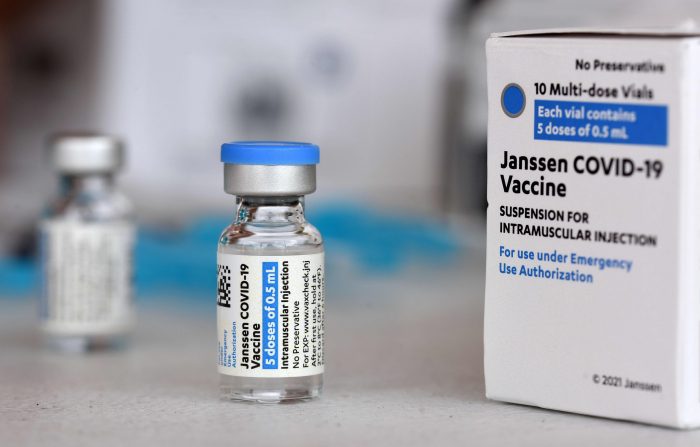
The Johnson & Johnson COVID-19 vaccine, also called Ad26.COV2.S, was developed by Janssen Pharmaceuticals of Johnson & Johnson.

It is a single-dose vaccine, which differentiates it from two-dose COVID-19 vaccines, such as the ones from Pfizer and Moderna.

In early 2021, the Food and Drug Administration (FDA) and, after a recommendation from the European Medicines Agency (EMA), the European Commission issued emergency and conditional authorization for the Johnson & Johnson vaccine for individuals aged 18 years and older. The vaccine has been approved for emergency use in 40 countries.

However, the FDA recommended a pause in the vaccine’s use, as a precautionary measure following a small number of rare blood clot incidents. At the same time, Johnson & Johnson delayed the distribution of the vaccine in Europe while the EMA conducted its own review.

On April 20, the company announced it would resume distribution of its vaccine in the European Union but with a safety warning, after the EMA concluded that the vaccine’s benefits continue to outweigh its risks.

After halting vaccination in the United States a few weeks ago, the FDA and the CDC recently recommended that vaccinations resume. They noted, however, that women under the age of 50 should be aware of the risk of rare blood clots.

Moreover, both agencies want women to know that alternative COVID-19 vaccines are available, for which there are currently no reports of rare blood clots.

The World Health Organization (WHO) has stated that, while it continues to monitor the safety responses from regulatory agencies, the vaccine is safe and effective against the severe risks of COVID-19.

How does the vaccine work?

The vaccine is categorized as a viral vector vaccine.

This vaccine form utilizes a different, harmless virus to deliver the genetic instructions for making the SARS-CoV-2 virus’ surface spike protein, which triggers the body’s immune response to produce antibodies. The vaccine does not contain the SARS-CoV-2 virus and cannot cause COVID-19.

The Johnson & Johnson vaccine has a 66% efficacy rate. While this is lower than the efficacy rates of the Pfizer or Moderna vaccines, it should be noted that the clinical trials of the Johnson & Johnson vaccine took place in different contexts — globally in regions where variants were more prevalent, and in the U.S. during a period of significant increase in COVID-19 cases.

The vaccine is around 85% effective at preventing severe disease from COVID-19, including hospitalization and death.

Common side effects

According to the FDA’s vaccine fact sheetTrusted Source, possible side effects include:

Allergies and anaphylaxis

As with other COVID-19 vaccines, the Johnson & Johnson vaccine has a low but potential risk of causing an allergic reaction.

A non-severe allergic reaction may include symptoms such as hives, swelling, rash, and respiratory problems.

A more severe, anaphylaxis response is much rarer. According to the FDA, a severe reaction can be characterized by:

An ingredient that has raised particular concerns about allergic response is polysorbate 80, a chemical that is structurally related to polyethylene glycol (PEG), found in Pfizer’s and Moderna’s mRNA vaccines, both of which have prompted similar worries.

While knowledge about the mechanisms of this allergy is limited, PEG and polysorbate allergies are extremely rare. For the Pfizer vaccine, an analysis of one patient who experienced PEG-related anaphylaxis showed that they had a history of allergic reactions to PEG-containing products.

An evaluation of vaccine allergies with PEG and polysorbate skin testing concluded that the usefulness of skin testing in predicting potential allergic responses remains unclear.

The CDC advises that anyone with a known allergy to any of the vaccine’s ingredients, including polysorbate 80, seek advice from a healthcare professional before receiving the COVID-19 vaccine.

People who have had a severe allergic reaction to any of the vaccine’s ingredients should not receiveTrusted Source this particular shot.

Controversy regarding blood clots

Similar to the AstraZeneca COVID-19 vaccine — Vaxzevria — the Johnson & Johnson vaccine was put on hold after eight cases of a rare blood clotting disorder, in combination with low blood platelet counts, were found in the vaccine recipients.

Most of these cases of thrombosis with thrombocytopenia were extremely rare types, including cerebral venous sinus thrombosis (CVST), which occurs in the brain.

All eight cases occurred within 21 days of vaccination in people under the age of 60. Also, the majority of the eight vaccine recipients who experienced these side effects were women.

The EMA’s safety assessment committee concluded on April 20 that the vaccine’s label should include blood clots as a potential rare side effect and state that the benefits of the vaccine outweigh these risks.

Authorities are uncertain what causes these cases of blood clotting. They suggest that it may be a triggered immune response similar to a rare condition of thrombocytopenia induced by heparin, which is a blood thinner.

CVST is rare in the general population, occurring at a rate of around 5 in 1 million people annually. Reports indicate that there have been eight cases among more than 7 million people who have received the Johnson & Johnson vaccine.

The benefits of receiving the vaccine outweigh the risk of blood clots and the risk of the complications that can come with COVID-19.

However, the pause is likely to have caused some hesitancy toward the vaccine and has slowed rollout.

“There is no doubt in my mind that there are groups for whom this vaccine is of benefit,” Dr. Paul Offit, director of the Vaccine Education Center at Children’s Hospital of Philadelphia, told The New York Times, “meaning that they’re more likely to get this vaccine than the other vaccines, whether it’s because of where they live, or because they’re homebound, or it’s hard to get a second dose.”

Dr. Offit warns that any vaccine hesitancy caused by this controversy would have “elevated a rare risk above a much more common risk,” that of developing COVID-19, which could place many people’s health in danger.

Researchers know that HDL, or good, cholesterol reduces inflammation. At healthy levels, HDL can also reduce the risk of stroke and heart attack.

So far, intervention trials using medications to improve or increase HDL levels have been unsuccessful. Also, some genetic research indicates that lifelong high or low levels of HDL do not seem to relate to cardiovascular outcomes, as expected.

That is why some cardiovascular risk prevention researchers are shifting their focus from circulating HDL levels to the actual functional ability of HDL.

Indeed, a research team from the Netherlands has studied the relationship between HDL’s ability to reduce inflammation and the risk of experiencing a first cardiovascular event.

“HDL are very complex particles with anti-atherosclerotic functions that are not reflected by measuring just the cholesterol quantity,” says senior study author Dr. Uwe J. F. Tietge, Ph.D., a professor and head of the Division of Clinical Chemistry at the Karolinska Institute in Stockholm, Sweden.

“Atherosclerosis [plaque buildup in the arteries] underlying cardiovascular disease is increasingly recognized as a disease with a strong inflammatory component, and a central biological function of HDL is to decrease inflammation.”

By analyzing data from 680 adults, the team found evidence to suggest that HDL’s ability to reduce inflammation is associated with a reduced risk of a cardiovascular event.

In the study, people who did not experience a cardiovascular event demonstrated higher anti-inflammatory HDL levels than participants who did experience a cardiovascular event.

“By using a novel research tool, our results provide strong support for the concept that plaque buildup in the arteries has an inflammatory component and that the biological properties of HDL particles have clinical relevance to cardiovascular disease risk prediction.” – Dr. Uwe J. F. Tietge, Ph.D.

The study appears in the journal Circulation, which is the American Heart Association’s (AHA) flagship journal. It received funding from the Netherlands Organization for Scientific Research and the Swedish Heart-Lung Foundation.

HDL: A natural anti-inflammatory

HDL is called good cholesterol because it picks up excess cholesterol in the blood and takes it back to the liver, which breaks it down and helps remove it from the body.

HDL can also reduce inflammation in the cells lining blood vessels. This occurs because HDL removes cholesterol stored in macrophage foam cells in atherosclerotic plaques and carries it to the liver.

Atherosclerosis occurs when plaque accumulates on the inner walls of arteries. Plaque is a substance made of cholesterol, calcium, fat, and other molecules.

As plaque deposits grow, they can gradually narrow the blood vessel, reducing blood flow and oxygen delivery to parts of the body or organs.

If atherosclerosis is severe enough, it can lead to major health concerns, including stroke, heart attack, and death.

When HDL removes cholesterol from foam cells in plaques, it helps reduce the size of the plaque. By reducing its size, it also reduces the amount of inflammation associated with the plaque.

Because of these factors, healthcare professionals typically include circulating HDL levels in many cardiovascular risk assessment tools.

HDL’s anti-inflammatory capacity

In the new study, the researchers included participants from a larger study: the PREVEND (Prevention of Renal and Vascular End Stage Disease) Study.

The PREVEND Study began in 1998 and is investigating the relationship between cardiovascular disease and kidney damage. The study has about 40,856 participants, all of whom are adults living in the city of Groningen in Northern Netherlands.

From this massive participant pool, the researchers behind the new study selected 680 participants. They excluded people who had experienced a cardiovascular event before the program tracking period.

The researchers used these 680 participants to create 340 case-control pairs of individuals. This meant that there were two groups: an experimental group of 340 people who experienced an initial cardiovascular event during the PREVEND tracking period and 340 people who did not.

The term “case-control pairs” refers to the fact that individuals from the experimental group were matched with someone from the control group who had the same sex, the same smoking status, and similar HDL levels. Each person in a pair had an age within 5 years of the other.

In the study, the researchers defined a cardiovascular event as experiencing a non-fatal or fatal heart attack, receiving a diagnosis of ischemic heart disease, or having surgery to open clogged coronary arteries.

The researchers extracted HDL from participant blood samples and assessed how much it was able to reduce inflammatory responses in endothelial cells, which line blood vessels.

They also measured levels of the participants’ C-reactive protein, which is a substance that increases in response to body-wide inflammation.

The researchers also assessed the participants’ cholesterol efflux capacity. This refers to how effectively their HDL removes cholesterol from cells similar to those in plaque.

After their analysis, the researchers found that HDL anti-inflammatory capacity was higher in people who did not experience cardiovascular events (31.6%) than in people who did (27%) during the follow-up period (1997–2009). For every 22% increase in HDL anti-inflammatory capacity, the risk of experiencing a heart event during the next 10 years reduced by 23%.

The participants’ HDL anti-inflammatory capacities were not related to circulating HDL cholesterol levels, cholesterol efflux efficiency, or C-reactive protein levels. The protective effect of increased HDL anti-inflammatory capacity was more powerful in women than men.

Improving cardiovascular risk prediction

Importantly, the team was also able to improve cardiovascular risk prediction by factoring HDL anti-inflammatory capacity into the Framingham Risk Score or replacing HDL levels with HDL anti-inflammatory capacity in the formula.

The Framingham Risk Score is a common tool that healthcare professionals use to assess the risk of developing coronary artery disease (CAD) during the next 10 years. It accounts for six CAD risk factors:

  • age
  • sex
  • total cholesterol levels
  • HDL levels
  • systolic blood pressure
  • smoking habits

The researchers write that their findings could have major clinical implications by providing healthcare professionals with more information to assess CAD risk.

If HDL inflammation capacity impacts CAD risk more than HDL levels, they could also help make currently used risk assessment tools more accurate.

Their findings could also encourage researchers to find medications to target or improve HDL inflammation capacity. This could offer healthcare professionals, and people at risk of CAD, a whole new avenue of preventive treatments.

Study limitations

Limitations in the study mean that scientists must now reproduce these findings in a much larger and far more diverse group of individuals.

The study included almost exclusively white participants. Just under 240 of the 340 case-control pairs in the study were male. The researchers also did not include information about stroke incidence in the study.

Furthermore, the study participants were genetically similar, coming from the same relatively small region of the world.

There are also no standardized methods of how to isolate HDL from plasma or how to test HDL functional abilities.

Still, despite these study drawbacks, the researchers are optimistic about their findings and what they could mean for millions of people going forward.

“The HDL cholesterol level is a good, established, simple, and cost efficient [cardiovascular disease] risk biomarker,” says Dr. Tietge.

“Our results, however, demonstrate that the anti-inflammatory capacity or assays looking at HDL function in general have the potential to provide clinically relevant information beyond the static HDL cholesterol measurements that are currently used.”

In a mouse study, scientists have shown that motor and non-motor symptoms of Parkinson’s disease are associated with two specific neural pathways.

The study paper, published in the journal Nature Neuroscience, opens the door to future research that may help develop interventions to treat the disease and its symptoms.

Parkinson’s disease

According to the National Institute on Aging (NIA), Parkinson’s disease is a progressive neurological condition that typically occurs in people aged 60 and older.

Parkinson’s primarily affects a person’s motor functions, that is, their ability to move their body in a coordinated manner. However, it can also have an impact on a person’s cognition and behavior, leading to mental health issues and problems with memory and attention.

According to the NIA, Parkinson’s occurs when neurons, or brain cells, die or become damaged. Cognitive and motor functions are affected by specific brain cells, and it is damage to these cells in particular that results in the symptoms of Parkinson’s.

Scientists do not know exactly why some people develop Parkinson’s. Research suggests it is likely to be due to a combination of hereditary and environmental factors, as well as the process of aging.

There is no known cure for Parkinson’s disease. As a consequence, treatments usually focus on relieving the symptoms caused by the condition.

To manage these symptoms, it is important to understand the structure of the parts of the brain that may be involved in the key symptoms of the disease.

However, the relationship between different neural circuits and the effects of Parkinson’s disease is still being explored.

Parkinson’s in the brain

In the present study, a team led by researchers from the University of California San Diego (UC San Diego) in La Jolla wanted to contribute to this knowledge.

They conducted a study in mice to understand the relationship between neural circuits in the brain and some of the functions that are typically affected by Parkinson’s disease, such as cognitive and motor abilities.

They looked in particular at the external globus pallidus (GPe) in the brains of mice, which previous research has linked to the motor symptoms of Parkinson’s.

The researchers used multiple methods to give a more detailed overview of this region of the mice’s brains, including electrophysiology, viral tracing, and behavioral experiments.

Importance for Parkinson’s therapy

The researchers identified two areas of the GPe and were able to relate these areas to motor skills and cognitive skills in the mice.

By manipulating neurons in these two parts of the brain, the scientists improved the mice’s locomotion and their reversal learning.

Dr. Byungkook Lim is an associate professor in the Neurobiology Section of the Division of Biological Sciences at UC San Diego and corresponding author of the study.

He explains, “our work demonstrates that the distinct neural circuitries in the basal ganglia are differentially involved in the motor and non-motor symptoms of Parkinsonian-like behaviors that occur at different stages of the disease.”

“This suggests that evaluation of the detailed circuit mechanisms is needed to fully understand the changes in [the] brain during the progression of [Parkinson’s disease] and could provide better therapeutic strategies for the treatment of [Parkinson’s disease].” – Dr. Byungkook Lim

The fact that specific neurons could be linked to particular changes in the brain regions of the mice means that it may be possible to develop new treatments for the symptoms of Parkinson’s.

In Dr. Lim’s words, “[s]elective manipulation of specific changes can rescue one type of symptom — without affecting other symptoms — of Parkinson’s disease.”

ferrantraite/Getty Images

Obesity can have severe effects on a person’s quality of life and lifespan. Research shows that it substantially increases the risk of a wide range of diseases, including type 2 diabetes, cardiovascular disease, and some cancers.

Despite the long-established health risks, however, recent decades have seen an inexorable rise in rates of obesity throughout the world.

One study found that between 1980 and 2015, the prevalence of obesity doubled in more than 70 countries and steadily increased in most others.

According to the World Health Organization (WHO), in 2016, more than 650 million adults had obesity. Overweight and obesity now claim the lives of more than 2.8 million people each year.

A major challenge for researchers is to distinguish the effects of genetics from those of lifestyle on the progression of obesity and its effects on health.

A team of researchers, led by two from the Obesity Research Unit at the University of Helsinki, in Finland, used an ingenious way to do this.

By studying 49 pairs of identical twins who did not share the same body mass index, or BMI, readings, they removed the effect of genes from the equation.

Identical, or monozygotic, twins have the same genetic makeup. They experience almost identical conditions in the womb and usually have very similar upbringings.

This means that the differences between the twins in the study arose from the influence of their environment or lifestyle as adults, rather than their genes or conditions during childhood.

The researchers have published their findings in the journal Cell Reports Medicine.

Biopsies of fat and muscle

First, the researchers took blood samples and biopsies of fat, or adipose, tissue and skeletal muscle tissue from the volunteers.

They then used a variety of molecular techniques to analyze the transcription of genes, production of proteins, and processing of metabolites in the two types of tissue.

A key finding was that the activity of mitochondria, the power plants of cells, was reduced in the muscle and adipose tissue of people with obesity.

The change was more marked in adipose than in muscle tissue.

Meanwhile, there was also increased inflammation in the tissues from twins with obesity, compared with their leaner siblings.

The changes in adipose tissue, but not muscle tissue, were associated with adverse health effects, including fatty liver disease and insulin resistance, conditions that have been linked to the development of diabetes.

Reduced power output

“If mitochondria […] are compared to the engine of a car, you could say that the power output decreases as weight increases,” explains senior author Prof. Kirsi Pietiläinen, also of the university’s Obesity Research Unit.

The researchers believe that these underperforming mitochondria produce more of the harmful reactive oxygen species, or free radicals, that stoke inflammation.

“A low-powered mitochondrial engine may also generate toxic exhaust fumes, which can cause a pro-inflammatory state in adipose tissue and, consequently, the onset of diseases associated with obesity,” explains Prof. Pietiläinen.

“What was surprising was that the mitochondrial pathways in muscle had no association with these adverse health effects,” she adds.

Another notable finding was that mitochondria in the adipose and muscle tissue of individuals with obesity were less efficient at breaking down a type of amino acid. These acids are the building blocks of proteins.

“This finding was of particular significance because the reduced breakdown of these amino acids and the resulting heightened concentration in blood have also been directly linked with prediabetic changes and the accumulation of liver fat in prior twin studies,” says Prof. Pietiläinen.

The researchers observe that in people with obesity, excessive nutrients may upset the control of catabolic and anabolic processes, reactions that break down and assemble organic molecules, respectively.

They write:

“We argue that, because of a high nutrition load, adipose tissue and skeletal muscle tissue no longer sufficiently shift between catabolic and anabolic reactions in acquired obesity. Consequently, the cells in these tissues increase their [internal] communication and activate emergency responses, such as inflammation.”

They acknowledge that the major limitation of their study was its cross-sectional design, meaning that it focused on a single point in time.

While the study identified associations between obesity and the various metabolic and health measures, it could not prove the existence of any causal relationships, as a result of this design

The miracle tree, Moringa, Moringa Oleifera, or the drumstick is a plant recognized for its medical properties. It is used to treat more than 300 conditions. All parts of the Moringa tree possess some medicinal or nutritional value” says Amrit Walia ( Amritpal Singh Walia ) Co-Founder of Pioneer Sustainable Agriculture PSA organic farm located in Nigeria.

One cup of fresh, chopped leaves contains protein, Vitamin B6, Vitamin C, Iron, Magnesium, Riboflavin, and Vitamin A.

In this article, you will be able to understand the five prime uses of Moringa leaf that clearly distinguished it as an extraordinary species:

1. Moringa Leaf is Rich in Antioxidants
Moringa acts to reduce high levels of free radicals which may cause oxidative stress leading to diseases like type 2 diabetes or heart disease.

The basic antioxidants in Moringa are beta-carotene and Vitamin C. The exact figure of the number of antioxidants in the Moringa herb is staggering.

A few antioxidants present include propyl gallate, tocopherols (Vitamin E), ascorbic acid, chlorogenic acid, hydroxyanisole (BHT) citric acid, vanillin, potassium, reduced glutathione, and more. Quercetin is also present and crucial in the regulation of blood pressure.

2. Moringa Leaf Regulates Blood Sugar
Moringa herb produces effects similar to isothiocyanates plant compounds able to regulate blood sugar.
Sufficient research shows a significant reduction in blood sugar after short-term use of the herb with meals.

For instance, it was discovered that a group of diabetics who added 50 grams of moringa leaves to a meal reduced the rise in blood sugar by 21%.

3. Moringa Leaf Works to Reduce Inflammation
If you incur injury and develop wounds, Moringa can help you to stop inflammation.

Moringa is stocked with nutrients and antioxidants which are found in other anti-inflammatory foods like tomatoes, fatty fish, olive oil, and certain fruits.

By fighting harmful free radicals, the herb prevents sustained inflammation which can result in chronic problems like cancer or heart disease.

4. Moringa Leaf Promotes Prostate Health and Cure ED
The leaves and seeds of the Moringa plant are rich sulfur-containing compounds called glucosinolates, which fight cancer in the body, more specifically, benign prostate hyperplasias.

The condition which is prone to attack elderly man is characterized by enlargement of the prostate which makes urination difficult. Moringa can reverse the effects of prostate cancer by significantly reducing prostate weight.

The herb also reduces levels of prostate-specific antigen which is produced by the prostate gland.

Moringa can also cure erectile dysfunction. Its leaves contain useful plant compounds called polyphenols which enhance blood flow by increasing nitric oxide production while simultaneously lowering blood pressure.

Conclusion
The points discussed above show how versatile Moringa leaf is in the fields of health and medical care. The plant can be used for detoxification and in the treatment of digestive disorders.

The list of other ailments this wonder plant can treat include the following – scurvy, kidney stones, Athlete’s foot, dandruff, epilepsy, paralysis, convulsions, splenomegaly, epilepsy, antispasmodic, joint pain, edema, rheumatism, arthritis, cardiac hypertrophy, coronary artery disease, myocardial infarction, heart failure, and many more.

Do you have any experience with the Moringa Leaf extracts? Kindly share with us in the comments below.

Scientists have offered a detailed view of the biochemical pathway surrounding Parkin — a protein that plays a key role in maintaining cellular energy.

The research, which appears in the journal Science Advances, may help scientists develop new treatments for Parkinson’s disease, type 2 diabetes, and cancer, which can all occur when Parkin is not functioning properly.

Mitophagy

Parkin’s central role is to remove damaged mitochondria, which are the power stations of the cell that are responsible for generating energy. Cellular stress can damage mitochondria.

Damaged or dysfunctional mitochondria tend to accumulate in the cell, and the body needs to clear them away using a process known as mitophagy. This process is essential for cell health and energy production.

Researchers have implicated mitochondrial dysfunction in neurodegenerative diseases such as Parkinson’s.

A key question for scientists has been how Parkin is able to respond to cellular stress so quickly. The signal for Parkin to do its work appeared to occur after it had already started.

Biochemical pathways

In the present study, the scientists gained a better perspective on the biochemical pathway that signals Parkin to clear away damaged mitochondria.

According to Prof. Reuben Shaw, director of the Salk Cancer Center in La Jolla, CA, and senior author of the study, “our findings represent the earliest step in Parkin’s alarm response that anyone has ever found by a long shot.”

“All the other known biochemical events happen at 1 hour; we have now found something that happens within 5 minutes.”

“Decoding this major step in the way cells dispose of defective mitochondria has implications for a number of diseases.”

Building on their prior research, the scientists searched for proteins that activate in response to the enzyme ULK1, which the enzyme AMPK triggers. They found that Parkin was a prime candidate.

The results were surprising as biochemical pathways are normally very complicated, involving up to 50 different proteins.

The scientists confirmed the findings using mass spectrometry. This process revealed how ULK1 was interacting with Parkin, offering a detailed view of how the various proteins work together.

It is this chain, from AMPK to ULK1 to Parkin, that accounts for the speed with which Parkin can react to cellular stress.

Future treatments?

By drawing a link between Parkin and AMPK, the research may aid in the development of new treatments for various diseases.

For Prof. Shaw, “the big takeaway […] is that metabolism and changes in the health of your mitochondria are critical in cancer, they are critical in diabetes, and they are critical in neurodegenerative diseases.”

“Our finding says that a diabetes drug that activates AMPK, which we previously showed can suppress cancer, may also help restore function in patients with neurodegenerative disease.”

“That is because the general mechanisms that underpin the health of the cells in our bodies are way more integrated than anyone could have ever imagined.”

According to the Centers for Disease Control and Prevention (CDC), the prevalence of obesity among adults in the United States increased from 30.5% in 1999–2000 to 42.4% in 2017–2018.

The prevalence of severe obesity almost doubled over the same period, from 4.7% to 9.2%.

One of the factors behind the steep rise in obesity rates may be the intensive marketing of high calorie, nutrient-poor food in restaurants, on screens, and on billboards.

Past research suggested that children with excess weight or obesity are more susceptible to food advertising, which may cause a cycle of weight gain and overeating.

Whether adults with obesity are also more prone to food marketing was unclear, however.

Researchers in Canada and France have found that women with obesity also appear to be more vulnerable to marketing — but that weight loss due to bariatric surgery can free them from the cycle.

If the scientists had found that marketing still holds sway over people after weight loss, this would imply an inbuilt vulnerability to overeating.

“That would mean people are endowed with unchangeable psychological characteristics that would always make them more responsive to marketing — which would make it very difficult to sustain a medically recommended weight,” says the study’s lead researcher Yann Cornil, Ph.D., from the University of British Columbia (UBC) in Vancouver, Canada.

“But one of the positive things is that after significant weight loss, people become less responsive to marketing, such that it is more sustainable to remain at a lower body mass index,” he adds.

The research, which was a collaboration between UBC and the Pitié-Salpêtrière University Hospital in Paris, France, appears in the Journal of Consumer Psychology.

Tests of susceptibility

The scientists compared the susceptibility to food marketing of three groups of women:

  • Group one: Comprising 73 women with severe obesity who were due to receive bariatric surgery (gastric bypass or band), which involves surgically altering the digestive system to promote weight loss.
  • Group two: Consisting of 41 women with moderate weight, who the scientists referred to as “lean controls.” The team matched these individuals to the first group in terms of their age, gender, income, employment, and marital status.
  • Group three: Comprising 29 women with obesity who were not in line to receive bariatric surgery or any other weight loss procedure.

The researchers tested the patients with obesity who were due for surgery before the procedure, then 3 and 12 months afterward.

They tested the second group twice, 6 months apart, and the third group only once.

The tests measured participants’ susceptibility to three common marketing strategies that portray food and portion sizes as more healthy than they actually are.

The first strategy, called the “health halo” effect, involves framing a product as natural or healthy.

The researchers asked participants to estimate the calorie content of 4 “healthy” snacks, including apple juice and granola bars, and 4 “indulgent” snacks, including a can of cola and chocolate bars.

In reality, the “healthy” snacks all contained more calories than the “indulgent snacks.”

The second marketing strategy involves downplaying portion sizes by renaming them, which tends to make people overeat.

The researchers asked participants to imagine choosing a portion of fries or a drink from a range of three options.

There were labels providing the actual size of each portion in grams or centiliters (cl), but these names either understated the sizes (as “mini,” “small,” and “medium”) or overstated them (as “medium,” “large,” and “extra-large.”)

The third marketing strategy involves omitting the smallest option in a size range, which can also lead to people choosing larger portions.

In this test, participants chose from a menu with 47, 65, and 95 cl size options, or from a menu with 24, 47, and 65 cl size variants.

The tests revealed that women with obesity were significantly more responsive than those with a moderate weight to the health halo and size labeling effects.

Crucially, however, women with obesity who underwent bariatric surgery were no more susceptible to these marketing ploys 12 months after the procedure than the control group with moderate weight.

Reward and motivation

In their paper, the scientists note neuroscientific research suggesting that brain regions involved in reward and motivation are more active in those with obesity.

They add that bariatric surgery may “rewire” the brain’s reward system in favor of more healthy food choices.

Alternatively, they write, the psychological impact of undergoing radical surgery to reduce their weight may motivate patients to eat more healthily.

At any rate, it remains unclear whether people who lose weight through other means, for example, through exercise or dieting, also become less responsive to food marketing.

The authors acknowledge that their study had some other limitations.

For example, they tested women with obesity only up to 12 months after surgery, so the durability of the effects beyond this point is unknown.

In addition, all the participants were adult French women. “Future research should therefore examine a larger and more diverse sample of people with obesity, including men and adolescents,” they write.

Stressful events — such as sitting an exam, giving a presentation, or attending a job interview — temporarily increase heart rate and blood pressure and dilate arteries.

This is a normal part of the body’s “fight-or-flight” response, but the lining of blood vessels, known as the endothelium, can take up to 90 minutes to recover after this kind of stress.

This may help to explain why a single episode of stress temporarily increases the risk of an acute cardiovascular event, such as a heart attack.

On the other hand, experts know that eating a diet rich in fruits and vegetables reduces a person’s risk of cardiovascular disease, heart attacks, and cardiovascular mortality.

Among the healthy nutrients found in fruits and vegetables are polyphenolic compounds known as flavanols. In the long term, these improve endothelial function and lower blood pressure.

The effects of flavanols during stress and in its immediate aftermath are unknown, however.

Scientists at the University of Birmingham in the United Kingdom have conducted the first study to investigate this question.

Their research suggests that drinking cocoa, which is rich in flavanols, in the hours before a stressful event can improve blood flow during the experience and help restore endothelial function in males aged under 45 years old afterward.

“Our findings are significant for everyday diet, given that the daily dosage administered could be achieved by consuming a variety of foods rich in flavanols — particularly apples, black grapes, blackberries, cherries, raspberries, pears, pulses, green tea, and unprocessed cocoa,” says senior author Dr. Catarina Rendeiro, Ph.D., of the university’s School of Sport, Exercise and Rehabilitation Sciences.

“This has important implications for measures to protect the blood vessels of those individuals who are more vulnerable to the effects of mental stress,” she adds.

The research, conducted by postgraduate student Rosalind Baynham, appears in the journal Nutrients.

Stress tests

The scientists recruited 30 healthy males between 18 and 45 years who underwent tests at the lab on two separate occasions at least 7 days apart.

The research involved them fasting for 12 hours before each session and abstaining from alcohol, vigorous exercise, and polyphenol-rich food and drink for 24 hours beforehand.

At the start of each visit, while the participants were relaxed, researchers assessed their “brachial flow-mediated dilatation,” which is a measure of endothelial function.

They also measured their blood pressure, heart rate, and blood flow.

On one of these visits, the participants drank regular cocoa, which is rich in flavanols, and on the other visit, they drank a special cocoa drink that was low in flavanols.

About 90 minutes later, they took a competitive test of mental arithmetic designed to elicit stress.

For example, during the test, the researchers filmed the participants, and they had to watch their performance on a screen. The researchers told the participants that “body language assessors” would assess them.

The arithmetic task got progressively faster, and the participants lost points whenever they got an answer wrong.

An experimenter in the room marked their responses and sounded an alarm when they made a mistake, or once every 10 answers if they did not.

At 30 and 90 minutes after the stress test, the researchers remeasured the participants’ endothelial function and blood pressure.

The results showed that 30 minutes after the stress test, all participants had impaired endothelial function. This measure remained significantly higher 90 minutes after the stress test.

However, participants who drank the high-flavanol cocoa saw smaller declines in endothelial function than those who consumed the low-flavanol cocoa.

In addition, high-flavanol cocoa increased blood flow during and after the test compared with low-flavanol cocoa.

Other cardiovascular and blood pressure measures were similar for both conditions.

Future investigations

The researchers note that the main limitation of their study was that it excluded females.

They report that, for the sake of consistency, they wanted to avoid the impact of hormonal fluctuations during the female menstrual cycle on the vascular changes under investigation.

This means that it is not possible to apply the outcomes of this study to females. Researchers remain unclear on the impact of flavanols on stress response in females.

However, existing evidence shows there are gender differences in how a body responds to stress. So, future studies should focus on females.

The researchers also write that a more in-depth investigation would include molecular measures of cardiovascular function. These should consist of nitric oxide, which is a signaling molecule that dilates blood vessels, and the stress hormone cortisol.

Finally, they write that future research should explore the effect of flavanol intake on stress responses in individuals at higher risk of cardiovascular diseases, such as high blood pressure, and people who experience mental stress, such as care workers.

Researchers have found that a 2-week increase in fiber intake can significantly alter a person’s gut microbiome, including increasing species of bacteria that break down fiber.

However, the quantity of short-chain fatty acids (SCFAs) did not increase. SCFAs are the result of bacteria breaking down fiber, and they have diverse roles within the body.

For instance, SCFAs are used as a source of energy for the cells of the colon and are involved in cell signaling. Some SCFAs also have anti-inflammatory properties and might influence insulin sensitivity and body weight.

The research, which appears in the journal mSystems, lays the groundwork for future studies to explore in more detail the relationship between fiber intake, gut bacteria, and SCFAs.

Benefits of fiber

Fiber plays an important role in human health. For example, a recent review of various meta-analyses found that people who eat the most fiber significantly reduce their chances of dying due to a cardiovascular event.

However, only 1 in 20 people in the United States consume the recommended amount of fiber.

According to Dr. Katrine Whiteson, associate professor of molecular biology and biochemistry, co-director of the University of California, Irvine (UCI) Microbiome Initiative, and co-author of the present study:

“The lack of fiber intake in the industrialized world is starving our gut microbes, with important health consequences that may be associated with increases in colorectal cancer, autoimmune diseases, and even decreased vaccine efficacy and response to cancer immunotherapy.”

The small intestine cannot digest fiber. Instead, according to the authors of the present study, it passes into the colon, where microbes are able to break the fiber down.

This process results in the production of SCFAs. Experts believe they are important for a range of factors affecting a person’s health.

The authors of the present study wanted to study the relationship between a short-term increase in dietary fiber, the makeup of the gut microbiome, and the presence of SCFAs.

Two-week diet

To investigate, the researchers conducted a study involving 26 undergraduate students enrolled on a biology course at UCI, as well as their instructors.

In week 1, participants ate their normal diet and provided three stool samples for analysis.

In week 2, the participants started a high fiber diet. They tracked their nutritional intake using a fitness app, aiming for 40 grams (g) of fiber each day. To assist, the researchers provided them with 10 meals each week that were high in fiber from a range of different plants.

In the third week, the participants were encouraged to increase their fiber intake to 50 g per day. During this week, participants provided another three stool samples.

According to graduate student Andrew Oliver, a teaching assistant for the course, “students raised their fiber intake by an average of 25 g per day, but the variability of pre-intervention fiber intake was substantial.”

“A few students had to go from nearly zero to 50 g daily by the end of the study. We all became a little obsessed with how much fiber was in the food we were eating.”

The researchers then analyzed the samples using DNA sequencing to identify the makeup of the bacteria. They used gas chromatography to measure SCFAs.

Microbiome composition

The researchers found that the composition of the participants’ gut microbiomes changed by around 8% following the dietary intervention.

This was largely due to increases in bacteria known for breaking down fiber, including BifidobacteriumBacteroides, and Prevotella.

However, the researchers saw no statistically significant increase in SCFAs. They speculate that this may be because stool samples do not accurately represent levels of SCFAs in the gut, which are primarily found in the cell walls of the intestines.

The researchers also suggest that the 2-week intervention may not have been long enough to see any difference in SCFAs.

According to Dr. Whiteson, “we hope to carry out longer dietary fiber interventions and study how fiber can support the gut microbiome and promote health.”

“At this time during a pandemic, when we need our immune health and healthy vaccine responses, we encourage everyone to think about the plant diversity of their diets and add some beans, berries, and avocados where they can.”