Exercise

1222982174 Image credit: klebercordeiro/Getty Images

Studies investigating the role of vitamin D in preventing or treating COVID-19 have drawn conflicting conclusions. But should a lack of evidence stop us from topping up our vitamin D levels as the Northern Hemisphere heads toward winter?

Most people know vitamin D as an essential vitamin for healthy bones and teeth. But researchers have attributed a host of other functions to the vitamin, and one of these is supporting the immune system.

A systematic review and meta-analysis from 2017 in BMJ drew on data from 25 randomized controlled trials to look at whether taking a vitamin D supplement could prevent acute respiratory tract infections.

The international research consortium, led by Prof. Adrian R. Martineau, from the Centre for Primary Care and Public Health and the Asthma UK Centre for Applied Research, at Queen Mary University of London, in the United Kingdom, looked at data from nearly 11,000 study participants.

Prof. Martineau and colleagues concluded that “Vitamin D supplementation was safe and it protected against acute respiratory tract infection overall.”

But does vitamin D have a part to play in COVID-19? By now, a number of studies have looked for links between the vitamin and the condition, and their findings have conflicted.

In this Special Feature, we investigate why some experts have suggested a link between COVID-19 and vitamin D, and we dig deep to explore how convincing the evidence from the latest studies really is.

We also discuss whether taking a vitamin D supplement can have realistic benefits, particularly for those in communities that have been hit the hardest by COVID-19.

Why vitamin D?

A number of experts have cited the 2017 study as circumstantial evidence that vitamin D may have a protective effect against COVID-19.

Their articles have appeared in journals such as The Lancet Diabetes & EndocrinologyBMJ NutritionPrevention & HealthMetabolism, and Aging Clinical and Experimental Research.

The common thread is that they highlight that adequate vitamin D levels may help our immune systems fight off the SARS-CoV-2 virus, as with other viruses that cause upper respiratory infections. People with vitamin D deficiency may, therefore, not be able to do this as effectively.

One aspect of this is that it provides an elegant excuse about why people from marginalized racial and ethnic groups have been disproportionately affected by COVID-19, as some scientists have suggested.

There is already evidence to suggest that people with darker skin tones who live in Northern latitudes have inadequate vitamin D levels.

To make vitamin D, our bodies convert a metabolite of cholesterol in our skin cells into an inactive form of vitamin D when we are exposed to sunlight, specifically to ultraviolet B (UVB) light. This inactive form then undergoes further chemical modification in the liver and kidneys.

The pigment melanin that gives our skin its color stops UVB light from reaching the cells. Hence, the darker a person’s skin, the more UVB light they need to make adequate levels of vitamin D from sunshine alone.

A study in the American Journal of Clinical Nutrition found that 17.5% of Black study participants in the United States were classed as being at risk of vitamin D deficiency, a figure nearly 8.5 times greater than the percentage of their white counterparts who were at risk of the deficiency.

Data from the past few months have shown that in the U.S. and the U.K., Black people are more likely to die if they have COVID-19 than white people.

Given the relationship between vitamin D and respiratory infections, it is perhaps not unsurprising that many people have suggested a tentative link between the vitamin and the disease.

So, let’s look at the studies that have sought to investigate this link in more detail.

Evidence so far

Back in June, the National Institute for Health and Care Excellence, in the U.K., reported that “There is no evidence to support taking vitamin D supplements to specifically prevent or treat COVID‑19.”

The organization based their statement on data from a number of published studies, all of which they deemed to contain a “very low quality of evidence.”

In August, a research team from the University of Glasgow, in the U.K., looked at the vitamin D levels of 341,484 participants in the U.K. Biobank health data repository. Of these, 656 had been to the hospital with COVID-19, and 203 had died.

Once the authors accounted for confounding factors, they concluded that there was no link between vitamin D levels and the likelihood of needing hospitalization for COVID-19 or dying from the disease.

The main limitation, the team noted, was that the vitamin D measurements had been taken roughly 10 years earlier.

Also in August, researchers in Spain reported the results of a small clinical study looking at intensive care unit (ICU) admissions and vitamin D supplementation.

The team gave one group of patients a supplementary high dose of calcifediol, a precursor molecule to vitamin D, in addition to a range of drugs to treat COVID-19. The other group did not receive calcifediol.

“Of [the] 50 patients treated with calcifediol, one required admission to the ICU (2%), while of [the] 26 untreated patients, 13 required admission (50%),” the researchers reported.

While these numbers seem impressive, the study was small and has several limitations. One is that the vitamin D levels of the participants were not measured before and during the study. There were also differences in confounding factors, such as other health conditions, between the two groups.

In addition, the study was open label, so both the researchers and the participants knew who had received vitamin D, which leaves room for bias.

Who tests positive for COVID-19?

A study published at the start of September in JAMA Network Open looked at the vitamin D levels of people who had received a COVID-19 test.

The research team, from the University of Chicago, in Illinois, used data from 4,314 people who had received a COVID-19 test at the university’s medical center between March 3 and April 10, 2020.

Looking at the medical records, they identified 489 individuals who had their vitamin D levels measured sometime in the year prior to the test, but not in the 14 days before it.

From this group, 71 people had received positive COVID-19 test results. Among them, 32 had vitamin D deficiency when their levels were last tested, and 39 did not have the deficiency.

The difference between these numbers did not reach statistical significance.

The team then used a model to predict how many people likely had vitamin D deficiency at the time of their COVID-19 tests, based on previous vitamin D tests and any information about subsequent vitamin D supplements.

When the researchers looked at positive COVID-19 tests in relation to predicted vitamin D status, their model showed that 21.6% of people who likely had the vitamin deficiency at the time of testing would receive positive COVID-19 test results. This figure was 12.2% among people without the deficiency.

While these data may indicate that vitamin D plays a role in the likelihood of receiving a positive COVID-19 test result, the researchers cautiously described the many limitations of their study.

In the paper, they note that “Randomized clinical trials of interventions to reduce vitamin D deficiency are needed to determine if those interventions could reduce COVID-19 incidence, including both broad population interventions and interventions among groups at increased risk of vitamin D deficiency and/or COVID-19.”

Also in September, the journal PLoS ONE published the findings of a large retrospective study conducted by researchers from Quest Diagnostics, in Secaucus, NJ, and the Boston University School of Medicine, in Massachusetts.

The team looked at data from 191,779 people with recorded COVID-19 test results and information about vitamin D levels from tests conducted in the preceding 12 months.

Their analysis showed that 8.1% of 27,870 people with adequate vitamin D levels had tested positive for a SARS-CoV-2 infection, while 12.5% of the 39,190 individuals with vitamin D deficiency had received positive results.

While the data are once again promising, there are a number of limitations. For example, the researchers used a model that the data did not fit particularly well.

Also, the vitamin D test results may not have accurately reflected each person’s vitamin D status at the time of their COVID-19 test. The team acknowledges that there may be other confounding factors that they did not control for.

In addition, it is worth noting that Quest Diagnostics sells a vitamin D test. And the only author of the study who is not directly affiliated with the company — Dr. Michael F. Holick, from Boston University — receives consulting fees from Quest Diagnostics and has authored a book that advocates vitamin D as a cure for common health problems.

Vitamin D and COVID-19 complications

Medical News Today recently reported on a study that looked at the vitamin D status of a group of patients who required hospital treatment for COVID-19.

The researchers found that only 32.8% of the 235 patients had vitamin D levels of at least 30 nanograms per milliliter, which they classed as sufficient. They also saw an association between sufficient vitamin D levels and having less severe COVID-19.

Although the study adds to the body of evidence that argues for a protective effect of vitamin D against COVID-19, it only included a small number of patients, and the researchers did not account for several potential confounding factors, including socioeconomic status, that could have had an impact on COVID-19 severity.

The authors themselves call for larger studies and randomized clinical trials to gain further insights.

Having a data set that is robust enough to account for a range of confounding factors will make the crucial difference between potential associations or correlations and a link that is underpinned by sound scientific data.

But do the effects really matter, when vitamin D can easily be supplemented?

Vitamin D supplements for all?

Writing in The Lancet Diabetes & Endocrinology, Prof. Martineau and Prof. Nita Gandhi Forouhi, of the University of Cambridge School of Clinical Medicine, in the U.K., recently suggested that it is worth taking a vitamin D supplement to ensure adequate levels while clinical studies into the link between vitamin D and COVID-19 are ongoing.

They explain:

“Pending results of such trials, it would seem uncontroversial to enthusiastically promote efforts to achieve reference nutrient intakes of vitamin D, which range from 400 [international units (IU) per day] in the U.K. to 600–800 IU per day in the U.S.A.”

“These are predicated on benefits of vitamin D for bone and muscle health, but there is a chance that their implementation might also reduce the impact of COVID-19 in populations where vitamin D deficiency is prevalent; there is nothing to lose from their implementation, and potentially much to gain,” the authors continue.

Many governments around the world have set recommended daily levels of the vitamin to ensure that people take in enough. This was true before COVID-19.

In the U.S., the National Institutes of Health (NIH) recommend that children older than 1 year and adults up to the age of 70 obtain 600 IU or 15 micrograms (mcg) of vitamin D each day. They advise people aged 71 or older to aim for 800 IU or 20 mcg.

The NIH recommend reaching these targets through a combination of the diet, sunlight exposure, and supplements.

Natural food sources of vitamin D include oily fish, beef liver, cheese, egg yolks, and mushrooms. Many breakfast cereals and milk and non-dairy alternatives are fortified with vitamin D, as are infant formulas.

In the U.K., Public Health England (PHE) recommend 400 IU or 10 mcg per day for people of all ages. Most people are able to get sufficient vitamin D from their diet and sunlight exposure in the spring and summer. This is not necessarily so during the rest of the year.

“Since it is difficult for people to meet the 10 [mcg] recommendation from consuming foods naturally containing or fortified with vitamin D, people should consider taking a daily supplement containing 10 [mcg] of vitamin D in autumn and winter,” PHE recommend.

They also say that people with little or no sunlight exposure due to work or personal circumstances and “Ethnic minority groups with dark skin from African, Afro-Caribbean, and South Asian backgrounds may not get enough vitamin D from sunlight in the summer and therefore should consider taking a supplement all year round.”

In light of the COVID-19 pandemic, the U.K. government is actively encouraging everyone to take a daily supplement of the vitamin, as many people may be spending more time indoors.

Of course, it makes sense for governments and public health bodies to recommend supplements for those struggling to get enough vitamin D.

But the consumption of dietary supplements is not ubiquitous, and there is variability among different racial and ethnic groups.

A 2016 study in JAMA that looked at trends in multivitamin consumption among U.S. adults found that in 2011–2012, 58% of non-Hispanic white study participants took multivitamins. For non-Hispanic Black participants the figure was 41% and for Mexican American participants it was 29%.

It is worth mentioning that excessive levels of vitamin D are toxic. “Vitamin D toxicity almost always occurs from overuse of supplements,” the NIH warn.

The upper daily limit of vitamin D for children aged 1–8, they report, is 63–75 mcg or 2,500–3,000 IU. For children aged 9 or over, teens, and adults, it is 100 mcg or 4,000 IU.

Vitamin D, COVID-19, and skin color

The risk of dying from COVID-19 is disproportionately high among people from marginalized ethnic and racial backgrounds.

In May, MNT reported on large study from the U.K. that found that preexisting conditions could not explain this increase in risk — but that there was a clear association with being a part of a racial or ethnic minority group or having experienced poverty.

Considering this data about COVID-19 risk and the fact that many people with darker skin in Northern climates do not have adequate vitamin D levels: Is the sunshine vitamin the reason that people from marginalized ethnic and racial groups are experiencing worse COVID-19 outcomes?

So far, the hypothesis remains just that.

Future scientific investigations into the suggested link between vitamin D status, COVID-19 outcomes, and skin color may provide clarity.

“Since African American and Hispanic populations in the U.S. have both high rates of vitamin D deficiency and bear a disproportionate burden of morbidity and mortality from COVID-19, they may be particularly important populations to engage in studies of whether vitamin D can reduce the incidence and burden of COVID-19,” the authors of the JAMA Network Open study discussed above note in their paper.

Yet vitamin D is likely only going to be one part of the complex puzzle that is COVID-19.

In a letter published in the Journal of Human Hypertension, a group from the U.S. and Argentina suggest that genetic susceptibility may be to blame. They point to a range of health conditions that affect African Americans more than white Americans.

“The usual explanation for these differences is the low socioeconomic status and educational levels, the social environment, lifestyle habits, and less access to healthcare services,” they write. “However, there are pieces of evidence that these non-favorable conditions are not enough, and there are other influential factors that may help [lead researchers] to a better approach to the real problem, like some genetic [factors].”

However, Dr. Winston Morgan, from the University of East London, in the U.K., has pointed to the lack of “evidence that the genes used to divide people into races are linked to how our immune system responds to viral infections,” in an opinion piece in The Guardian.

Instead, there is mounting evidence that structural racism is a crucial factor in why marginalized communities are harder hit by COVID-19.

In an exclusive opinion piece for MNT, Dr. Morgan discussed the outcomes of a recent PHE review into why COVID-19 disproportionately affects people from marginalized racial and ethnic groups.

He notes that the review’s recommendations focus on the need to address structural problems in health outcome disparities.

Vitamin D does get a mention. The review’s authors highlight the need for “further evidence as a matter of urgency” in order to deepen our understanding of why people of color are disproportionately experiencing negative outcomes of COVID-19.

In an interview with MNT, Assistant Prof. Tiffany Green, from the University of Wisconsin-Madison School of Medicine and Public Health, explained that “Those of us who work in the health disparities space are saddened but not surprised at the race-based disparities that the COVID-19 crisis has brought to light.”

She pointed to the “racialized class and occupational structures of the U.S.” as a major factor that contributes to who is exposed to the SARS-CoV-2 virus.

To conclude, it makes sense to look after our vitamin D levels as part of our general health, and by extension our ability to fight off infections. But science rarely has easy answers.

In order to navigate our way out of the COVID-19 pandemic, we would be better served if we were able to accept that we are up against a complex interplay of societal and immunological factors.

113174398 Image credit: Dougal Waters/Getty Images

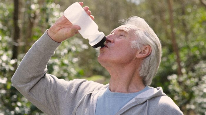
A new study confirms that older men may lack the bodily cues that help younger men remain hydrated.

Smart thermostats have nothing on our hypothalamus. This is the gland that helps us maintain a healthy body temperature.

When we get too hot, the hypothalamus causes our skin to produce sweat that cools us down as it evaporates. We then become thirsty, and we should drink to replace the water that we lost through sweating.

However, if we sweat too much or do not drink water to replenish our fluids, we can become dehydrated.

Without enough water in the body, we lose the ability to cool ourselves with sweat, and the body can overheat. This increases our chance of experiencing heatstroke and other heat-related damage to our bodies.

As we age, the efficiency of our temperature regulation system declines. Although most research into the effects of dehydration focuses on young adults, a new study in The Journal of Physiology examines its role in the health of older adults.

Older adults may not feel as thirsty as young people and should take care to hydrate when they work or exercise and when the weather is hot.

Dehydration changes as we age

Researchers from the Human and Environmental Physiology Research Unit at the University of Ottawa in Canada explored the paradoxical risk associated with dehydration later in life.

On the one hand, during exercise, dehydration in older adults does not lead as readily to an increase in body temperature through a reduction in heat loss as it does in younger people.

Although this may seem to be a good thing, the lack of sweat and thirst means that the person loses important cues that suggest that it is time to rehydrate.

Without drinking enough water, dehydration in older adults may persist and quietly increase to dangerous levels.

Blood salinity

Scientists have suggested that the reason that older adults feel less thirsty is due to a reduced ability to detect and respond to the level of salt in their blood.

When the balance between water and salt in the blood tips toward salinity, the body of a younger adult responds with feelings of thirst.

The researchers wondered if the same reduced ability to track blood salinity, or “osmolality,” that reduces sensations of thirst may also be the driver behind the less extreme response to dehydration in older adults.

Ten younger men (18–30 years old) and 10 older men (54–67 years old) participated in exercise heat stress tests. The researchers asked them to abstain from consuming alcohol and engaging in strenuous exercise for 24 hours before each session. They also asked them to drink 500 milliliters of water the night before the experiments.

After screening, the men took part in two exercise sessions placed a week apart. At the start of each exercise session, the participants received an intravenous saline solution to increase blood osmolality before entering a heated, whole-body direct-air calorimeter for 1 hour of stationary cycling.

The calorimeter measured the participants’ whole-body evaporative and dry heat loss, and other measurements tracked a range of body indicators of temperature and rate of heat loss.

Analysis of these data revealed a substantive difference in the regulation of body temperature between the younger and older men.

The researchers found that for older men, an increase in blood salinity did not trigger the body’s responses to dehydration as it did in the younger men.

An incomplete picture

Further research will be necessary to help scientists gain a full understanding of heat regulation in older adults.

As first study author Robert Meade says, “While our research design allowed us to test the independent effect of osmolality on heat loss, the effect of reduced blood volume (termed hypovolemia) on sweating in older adults is currently unknown.”

Because the study explored the effects of blood osmolality in physically active participants without any known chronic conditions, it is unclear whether or not the same finding would apply to older adults with common age-related conditions such as type 2 diabetes.

However, Meade concludes:

“Given that common age-related chronic health conditions such as type 2 diabetes are associated with less efficient regulation of body temperature and hydration status, future research should be conducted to see whether our findings translate to or are exaggerated in those populations.”

1094048132 Image credit: Cavan Images/Getty Images

A study finds that 10 minutes of massage or relaxation can activate the body’s system for overcoming stress.

The damaging effects of stress are well-known, but fortunately, our bodies have a built-in system for managing and recovering from it. This system is called the parasympathetic nervous system (PSNS).

While there is plenty of anecdotal evidence that taking time to relax — especially when it involves massage — can activate the PSNS, the new study by psychologists at the University of Konstanz in Germany has scientifically measured and confirmed this effect.

In their paper, the researchers conclude that short periods of relaxation may be psychologically and physiologically regenerative and that the effect is even more pronounced with massage.

The senior author of the study is Prof. Jens Pruessner of the university’s Neuropsychology lab, who is a member of the Cluster of Excellence “Centre for the Advanced Study of Collective Behaviour.” He explains the importance of the new research:

“To get a better handle on the negative effects of stress, we need to understand its opposite — relaxation. Relaxation therapies show great promise as a holistic way to treat stress, but more systematic scientific appraisal of these methods is needed.”

The study appears in the September 2020 issue of Scientific Reports.

The study

For their study, the team divided the participants into three groups.

The first group received 10-minute head-and-neck massages with a moderate pressure intended to stimulate the PSNS’s vagus nerve. This nerve contains some 75% of the PSNS nerve fibers, branching out to the many organs in the body with which the system interacts.

The second group of individuals received much softer 10-minute neck-and-shoulder massages as a means of determining the PSNS-activating effect of simple tactile contact.

A third control group simply sat at a table relaxing for 10 minutes.

The researchers used both physiological and psychological measurements to evaluate the degree to which each intervention, or lack of, had activated the participants’ PSNS.

Neuropsychology doctoral student Maria Meier led the team, who assessed the tests’ physiological effect by measuring the participants’ heart rate, as well as their heart rate variability (HRV). HRV is a measurement of variations in the time intervals between heartbeats.

For example, when the body is in fight-or-flight mode, there is very little variation because the heart beats quickly at a steady rate. This will provide a low HRV value. When the body is relaxed, a greater degree of variation occurs, resulting in a higher HRV.

All of the participants had significantly higher HRV levels afterward. However, the most dramatic increases in HRV belonged to those who had received massages. The type of massage did not matter.

Simple tactile contact proved just as effective for helping an individual relax as a massage designed specifically to activate the PSNS.

Psychologically, all participants reported feeling less stressed and more relaxed after the tests.

Overall, the experiments confirmed that simply taking a few moments to relax can help a person manage stress. Adding a relaxing massage does even more to activate the PSNS and alleviate the physical and mental effects of stress.

Stress management

Meier concludes: “We are very encouraged by the findings that short periods of disengagement are enough to relax not just the mind but also the body. You don’t need a professional treatment in order to relax. Having somebody gently stroke your shoulders, or even just resting your head on the table for 10 minutes, is an effective way to boost your body’s physiological engine of relaxation.”

Equally important as the study’s finding is the development of a system for objectively evaluating relaxation therapies. With experts often citing stress as the driver of diseases such as depression, a reliable means of validating relaxation techniques clearly has value.

Says Meier, “Massage, being such a commonly used relaxation therapy, was our first study. Our next step is to test if other short interventions, like breathing exercises and meditation, show similar psychological and physiological relaxation results.”

1187813695

Data spanning more than 11 years suggest that testosterone injections could be a novel treatment for obesity in men. The results show that long-term testosterone therapy may be comparable to weight loss surgery, with a lower risk of complications.

Over 42% of adults in the United States have obesity, according to the Centers for Disease Control and Prevention (CDC). Obesity has links to several chronic health conditions, including heart disease and type 2 diabetes.

Recent findings that obesity may also worsen outcomes in COVID-19 have encouraged some governments to create entirely new public health strategies to encourage people to lose weight.

However, obesity is a complex issue with both medical and social causes, and achieving lasting weight loss can be challenging for many people. This means researchers are looking for new strategies to help treat obesity — beyond merely cutting calories.

Data recently presented at the virtual European and International Congress on Obesity support the use of testosterone therapy to treat men with obesity.

Long-term testosterone treatment reduced body weight by 20% on average.

11 years of data

The pharmaceutical company Bayer and Gulf Medical University in the United Arab Emirates led the research, using 11 years’ worth of data.

The researchers collected data since 2004 of 471 men with functional hypogonadism, or low testosterone production, and obesity from a German urological practice.

Around 58% of the men received an injection of testosterone every 3 months for the duration of the study, while the remainder chose not to have the treatment and therefore acted as controls. The average age of the participants was 61.57.

Medical staff administered and documented all injections at a doctor’s office, which assures that all participants received the treatment in a consistent manner. No participants dropped out of the study.

20% bodyweight reduction

The men who received testosterone lost on average 23 kilograms (kg) (equivalent to 20% body weight) during the study period, while those who did not receive treatment gained an average of 6 kg.

Body mass index (BMI) correspondingly decreased by an average of 7.6 points in those who received testosterone therapy, compared with an increase of 2 points in the control group.

Waist circumference, which is a risk factor for cardiometabolic disease, decreased by an average of 13 centimeters (cm) in the treatment group, compared with a 7 cm increase in the control group.

The testosterone-treated men also had less internal (visceral) fat by the end of the study period. They may have had a lower risk of cardiovascular disease than those who did not receive treatment.

Overall, 28% of men in the control group had a heart attack, and 27.2% had a stroke during the study period. There were no major cardiovascular events in the men who received testosterone therapy.

Likewise, while more than 20% of the control group developed type 2 diabetes during the study period, nobody in the treatment group developed the condition.

Commenting on the results, Farid Saad of Bayer said: “Long-term testosterone therapy in hypogonadal men resulted in profound and sustained […] weight loss, which may have contributed to reductions in mortality and cardiovascular events.”

An alternative to surgery?

The researchers also presented data specific to men who were eligible for bariatric surgery. This is a surgical treatment for obesity, which encompasses gastric band, gastric bypass, and gastric sleeve surgery.

Rates of bariatric surgery are on the rise in the U.S., with more than 250,000 people undergoing weight loss surgery in 2018 alone.

Although bariatric surgery is a proven means of achieving weight loss, there are serious risks associated with the surgery, which does not always have positive outcomes.

This part of the study included 76 men with class 3 obesity (a BMI of 40 or above), making them eligible for bariatric surgery. Of these, 59 received testosterone treatment and lost 30 kg on average.

The BMI of the men also reduced by an average of 10 points, which could be enough to take them out of the highest obesity class, provided their BMI was less than 50 to begin with.

According to Saad, these results suggest testosterone therapy could be as effective as surgery for weight loss — but without the risk of serious complications.

“We believe testosterone therapy should be discussed with patients as an alternative to surgery and should be considered for male patients who cannot undergo surgery.” – Farid Saad, Consultant in Medical Affairs Andrology, Bayer AG

This study is based on data exclusively from men, and all participants had clinically low testosterone levels. Scientists need to conduct further studies to validate the use of this approach in other populations.

535150115

A study of older adults in the United Kingdom finds that people who are lonely are more likely to develop type 2 diabetes, independent of other risk factors such as smoking, alcohol consumption, and weight.

Loneliness, in which a person’s social needs are not met, may be on the rise. A recent report found that almost half of people in the United States sometimes or always feel alone.

Loneliness is even more common among younger generations, with almost 80% of Gen Z and more than 70% of millennials experiencing this feeling.

Some believe that technology may play a part in feelings of loneliness among younger generations, with social media and other forms of online communication increasingly replacing genuine human connection.

Beyond the negative emotional impact of feeling isolated, loneliness is also a major risk to physical health. Research has associated loneliness with coronary heart disease and found that loneliness may be a greater threat to health than obesity.

One study even suggested that people who are lonely have a higher mortality risk than individuals who do not feel alone.

A new study from Kings College London in the U.K. adds to the list of health concerns associated with loneliness, finding that people who are lonely may be more likely to develop type 2 diabetes.

The researchers found that loneliness was a significant predictor of diabetes. This finding held when they took account of potential confounding factors, such as age, sex, ethnicity, wealth, smoking, physical activity, body weight, alcohol consumption, hypertension, and cardiovascular disease.

The findings appear in the journal Diabetologia.

The cohort

The study, which is the first to find an association between loneliness and type 2 diabetes, is based on data from more than 4,000 people aged 50 years and above, with an average age of 65 years. The collection of the data took place during the English Longitudinal Study of Ageing.

At the start of the current study, none of the participants had diabetes, and they all had blood glucose levels within a healthy range.

During a follow-up period of 12 years, 264 people in the study (roughly 6% of the sample) developed type 2 diabetes.

The researchers found that the level of loneliness that people experienced at the start of the study was a significant predictor of who would go on to develop diabetes.

Loneliness predicts diabetes onset

The assessment of loneliness occurred at the start of the study using a scale that a psychologist at the University of California, Los Angeles, developed. The scale requires people to rate questionnaire items such as “How often do you feel that you lack companionship?” and “How often do you feel part of a group of friends?”

The researchers found a significant association between loneliness and the onset of type 2 diabetes, even when they controlled for confounding factors, including smoking, alcohol consumption, weight, blood pressure, and cardiovascular disease.

The association was also independent of mental health factors, such as depression and whether a person lived alone.

“The study also demonstrates a clear distinction between loneliness and social isolation, in that isolation or living alone does not predict type 2 diabetes, whereas loneliness, which is defined by a person’s quality of relationships, does,” explains lead author Dr. Ruth Hackett.

This finding highlights the importance of the quality of human interactions that a person has, rather than the quantity.

Stress-related mechanism?

Although the reason for this association is not yet clear, the researchers suggest that it could be related to how the body manages stress.

Previous research has shown, for example, that loneliness is associated with changes to levels of the stress hormone cortisol, which plays a role in diabetes.

“If the feeling of loneliness becomes chronic, then every day you’re stimulating the stress system, and over time, that leads to wear and tear on your body, and those negative changes in stress-related biology may be linked to type 2 diabetes development,” explains Dr. Hackett.

However, it is important to note that this is currently only a hypothesis. Although this study provides a correlation between loneliness and type 2 diabetes, it does not show a causative link between the two factors.

Other limitations of the study include the fact that there was only one measurement of loneliness during the study. Also, the data on type 2 diabetes were based on self-reporting, rather than objective medical records.

The authors also note that the overall strength of the association between the two factors was small. Nevertheless, the study highlights loneliness as a potential risk factor for type 2 diabetes and provides a basis for future studies to investigate this connection in more detail.

Scientists have found associations between fungi living in the gut and mild cognitive impairment, which can lead to Alzheimer’s disease. They suggest that a ketogenic diet could help prevent the disease by creating a more healthful balance of microorganisms in the gut.

Alzheimer’s Association report that 15–20% of people over 65 experience mild cognitive impairment (MCI), which involves a decline in memory and the ability to think clearly.

Doctors do not consider MCI to be a form of dementia because people who have it are able to function relatively well and live independently. However, MCI is associated with an increased risk of Alzheimer’s disease or another form of dementia later in life.

Developing preventive strategies that can be initiated early is therefore a priority. But despite decades of research, no therapies have been shown to reverse or prevent the brain changes seen in Alzheimer’s.

One potential strategy that scientists have begun to explore involves modifying the diet to influence the microbial communities in the gut.

There is an intimate relationship between the gut microbiome and the central nervous system, with recent research suggesting associations between particular bacterial communities and neurological disorders, including MCI, dementia, and Alzheimer’s.

In one recent study, scientists at the Wake Forest School of Medicine, in Winston-Salem, NC, found a distinctive gut bacterial “signature” in people with MCI.

They also discovered that a diet called the modified Mediterranean-style ketogenic diet altered bacterial communities in the guts of volunteers and reduced biomarkers of Alzheimer’s disease in the cerebrospinal fluid of those with MCI.

This diet contains a limited number of carbohydrates and increased amounts of fats. These are primarily mono- and polyunsaturated fats that come from olive oil and fish.

ketogenic, or “keto”, diet contains very few carbohydrates, which the body uses as fuel. When short on carbs, the body starts to break down its fat reserves to produce molecules called ketones, as an alternative source of energy.

The same group of researchers now reports similar associations between the communities of fungi in the gut — collectively called the mycobiome — the person’s diet, and their risk of MCI.

They report their findings in the latest issue of the journal EBioMedicine.

Fungi and brain health

“Although we do not fully understand how these fungi contribute to Alzheimer’s disease, this is the first study of its kind to reveal their role in our mental health, which we hope will ignite thinking in the scientific community to develop better understanding of them in relation to Alzheimer’s disease,” says principal investigator Hariom Yadav, Ph.D., an assistant professor of molecular medicine at Wake Forest.

“It also indicates that dietary habits such as eating a ketogenic diet can reduce harmful fungi in the gut, which might help in reducing Alzheimer’s disease processes in the brain,” he explains.

The researchers randomly assigned 17 older adults to eat either a Mediterranean-style ketogenic diet (MMKD) for 6 weeks or the American Heart Association diet, which is relatively low in fat and high in carbohydrates.

Eleven of the participants had MCI and six had no cognitive issues. Their average age was 65 years.

The team provided each participant assigned to the MMKD with 2 liters of extra virgin olive oil and encouraged them to eat fish, lean meats, and nutrient-rich foods.

After a 6-week “washout” period during which the participants maintained their regular diets, they switched to their study-assigned diets for 6 weeks.

The scientists monitored changes in the participants’ mycobiomes by analyzing fecal samples and changes in Alzheimer’s biomarkers by assessing samples of cerebrospinal fluid.

Fungal signature and diet

At the start of the study, the mycobiomes of participants with MCI had a distinctive signature, containing more of certain types of fungi and fewer of others, compared with those of the other participants.

As expected, MCI was associated with a high-risk profile of Alzheimer’s biomarkers in cerebrospinal fluid. In keeping with previous findings, following an MMKD appeared to improve this risk profile.

Interestingly, individuals with MCI initially had less fungal diversity in their guts, compared with the healthy participants, but eating an MMKD seemed to restore the diversity.

The researchers note that changes in the populations of gut bacteria of individuals with MCI who ate an MMKD — which they had reported in their previous study — may have had knock-on effects on the fungi.

Some species of bacteria produce substances known to suppress the growth of certain fungi. The authors report that this may explain why eating an MMKD suppressed a genus of fungi called Candida in the guts of participants with MCI.

Candida has been implicated in a range of inflammatory diseases of the gut, including Crohn’s disease and ulcerative colitis.

Inflammation is now believed to play an important role in Alzheimer’s disease, as well. By suppressing Candida and reducing inflammation, say the researchers, eating an MMKD may help reduce the risk of Alzheimer’s.

Study limitations

The authors acknowledge that their study had some key limitations. First, while the researchers advised people to eat certain diets, they did not monitor the participants for this.

It was also a pilot study with a small sample size, making it impossible to account for other variables that might affect Alzheimer’s risk and the population of the gut’s microorganisms, such as lifestyle, gender, and ethnicity.

Moreover, the unique signature of gut fungi and bacteria found in individuals with MCI may be a result of the health issue, rather than one of its causes.

Finally, the changes in the mycobiome observed in the participants assigned the keto diet may have been transient, resulting from fungi in the food.

Eating in the evening is associated with a higher intake of calories, as well as lower quality food, according to a new study.

Maintaining a healthful diet is associated with how late in the day people consume most of their food, according to research presented at the European and International Conference on Obesity (ECOICO 2020).

The study found that people who consume most of their calories in the evening tend to consume more of them and have a lower quality diet.

The study’s aim was to explore the connection between the evening consumption of calories — the measure of energy intake (EI) — and diet quality. Judith Baird, a researcher from the Nutrition Innovation Centre for Food and Health at Ulster University in Northern Ireland, United Kingdom, led the study.

Hunger rhythms

Previous studies have found that hunger follows a daily rhythm and that this rhythm is, in some ways, not what people might expect. Although people typically cease eating during an extended period of sleep, they break that fast with what is often the smallest meal of the day.

Meanwhile, hunger tends to be strongest late in the day, peaking at about 8:00 p.m., after most people have completed the majority of their daily activities.

EI consumption naturally tends to be a response to hunger, and other research has investigated the effect of meal timing on metabolism and other bodily processes. The new study, however, looks at its implications for the quantity and quality of food that people consume.

Data used in the study

Beginning in 2008, the U.K.’s National Diet and Nutrition Survey (NDNS) captured detailed information regarding food consumption, nutrient intake, and nutritional status for individuals over the age of 18 months. Each year, the survey collected responses from a representative sample of 1,000 people. Baird and her colleagues analyzed data from 1,177 adults who participated in the survey from 2012 through 2017.

Overall, the researchers found that the participants were, on average, consuming nearly 40% (39.8%) of their daily EI after 6:00 p.m.

Looking at the data more closely, the researchers divided people into quartiles according to the proportion of their daily EI that they consumed after 6:00 p.m. The people in the lowest quartile consumed less than 31.4% of their EI in the evening, while those in the highest quartile ate more than 48.6% during evening hours.

What the data say

The researchers detected two significant trends in the data. First, the study found that eating later affected the total EI for the day.

People who consumed most of their daily EI earlier tended to eat fewer calories over the course of a day.

The findings also suggested that meal timing affects the nutritional quality of food. Baird and her colleagues assessed individuals’ diets as they had reported them in the food diaries that they had supplied to the NDNS. To do this, they consulted the rankings listed in the Nutrient-Rich Food Index. The index rates foods according to their ratio of important nutrients to calorie value.

People who consumed more of their calories during the evening tended to have significantly poorer quality diets.

“Our results suggest that consuming a lower proportion of EI in the evening may be associated with a lower daily energy intake, while consuming a greater proportion of energy intake in the evening may be associated with a lower diet quality score. – The study authors

The study authors present their insights as just one facet of a deeper understanding of the effect of a person’s daily food rhythms and the amount and quality of food that they consume. They conclude:

“Timing of energy intake may be an important modifiable behavior to consider in future nutritional interventions. Further analysis is now needed to examine whether the distribution of energy intake and/or the types of food consumed in the evening are associated with measures of body composition and cardiometabolic health.”

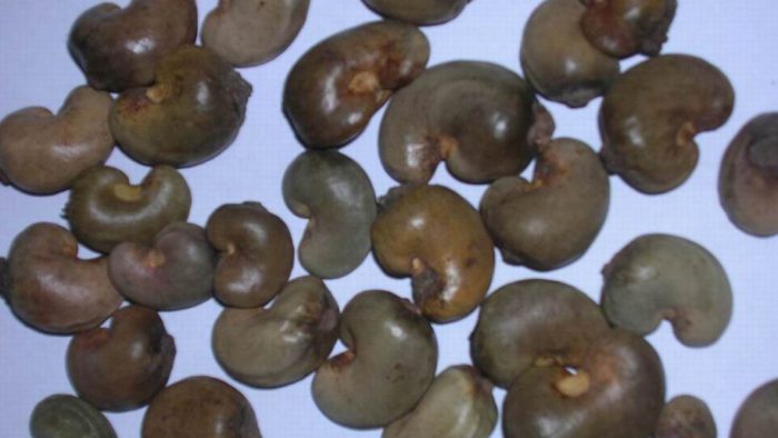
In laboratory experiments, a chemical compound found in the shell of the cashew nut promotes the repair of myelin, a team from Vanderbilt University Medical Center reports Tuesday in the Proceedings of the National Academy of Sciences.

Myelin is a protective sheath surrounding nerves. Damage to this covering — demyelination — is a hallmark of multiple sclerosis and related diseases of the central nervous system.

“We see this as an exciting finding, suggesting a new avenue in the search for therapies to correct the ravages of MS and other demyelinating diseases,” said Dr. Subramaniam Sriram, paper’s senior author, William C. Weaver III Professor of Neurology and chief of the Division of Neuroimmunology.

Previous work led by Sriram showed that a protein called interleukin 33, or IL-33, induced myelin formation. IL-33 is, among other things, an immune response regulator, and multiple sclerosis is an autoimmune disorder.

The cashew shell compound is called anacardic acid. Sriram and team grew interested in it because it is known to inhibit an enzyme involved in gene expression called histone acetyltransferase, or HAT, and the team had discovered that whatever inhibits HAT induces production of IL-33.

The report includes a range of new findings that point to the potential therapeutic use of anacardic acid for demyelinating diseases:
*In vitro, the addition of the compound to rat cells most responsible for myelination — oligodendrocyte precursor cells, or OPCs — spurred induction of IL-33 and rapidly increased the expression of myelin genes and proteins, including dose-dependent increases in myelin basic protein;

*In two animal models of demyelination, treatment with the compound increased the relative presence of IL-33-expressing OPCs and led to reduced paralysis;

*In an animal model of demyelination treated with the compound, dissection and electron microscopy showed dose-dependent increases in myelination.

“These are striking results that clearly urge further study of anarcardic acid for demyelinating diseases,” Sriram said.

If your cholesterol level has crept up over the years, you may wonder whether changing your diet can help. Ideally, your total cholesterol value should be 200 milligrammes per deciliter (mg/dL) or lower. But it is the harmful Low-Density Lipo-protein (LDL) ‘bad’ cholesterol value that experts worry about the most. Excess LDL builds up on artery walls and triggers a release of inflammatory substances that boost heart attack risk.

“To prevent heart disease, your LDL should be 100 mg/dL or lower,” says Dr. Jorge Plutzky, director of preventive cardiology at Harvard-affiliated Brigham and Women’s Hospital. But many Americans have LDL values that are less than optimal (100 to 129 mg/dL) or borderline high (130 to 159 mg/dL).

If you fall into either of those categories, you may be able to nudge down your LDL to a healthier level by changing what you eat, particularly if your current diet could use some improvement. However, most people with higher LDL values likely will also need to take a cholesterol-lowering drug, such as a statin, says Dr. Plutzky.

Dietary directives
Avoiding foods that are high in cholesterol isn’t the best way to lower your LDL. Your overall diet — especially the types of fats and carbohydrates you eat — has the most impact on your blood cholesterol values. “As the American Heart Association has noted, you’ll get the biggest bang for your buck by lowering saturated fat and replacing it with unsaturated fat,” says registered dietitian Kathy McManus, director of the Department of Nutrition at Brigham and Women’s Hospital.

That means avoiding meat, cheese, and other high-fat dairy products such as butter, half-and-half, and ice cream. Equally important is replacing those calories with healthy, unsaturated fats (such as those found in vegetable oils, avocados, and fatty fish) rather than refined carbohydrates such as white bread, pasta, and white rice. Unlike healthy fats, these starchy foods aren’t very filling, and they can trigger overeating and weight gain.

The other big problem with refined carbs: They are woefully low in fibre, which helps flush cholesterol out of the body.

The fibre factor
Your body can’t break down fiber, so it passes through your body undigested. It comes in two varieties: insoluble and soluble. Fiber-containing foods usually feature a mix of the two.

Insoluble fibre does not dissolve in water. While it doesn’t directly lower LDL, this form of fiber fills you up, crowding other cholesterol-raising foods out of your diet and helping to promote weight loss.

Soluble fibre dissolves in water, creating a gel. This gel traps some of the cholesterol in your body, so it’s eliminated as waste instead of entering your arteries.

Soluble fibre also binds to bile acids, which carry fats from your small intestine into the large intestine for excretion. This triggers your liver to create more bile acids — a process that requires cholesterol. If the liver doesn’t have enough cholesterol, it draws more from the bloodstream, which in turn lowers your circulating LDL.

Finally, certain soluble fibres (called oligosaccharides) are fermented into short-chain fatty acids in the gut. These fatty acids may also inhibit cholesterol production.

The “best” foods
The following 11 foods are good sources of fibre or unsaturated fat (or both). But they’re not in any particular order and are simply suggestions. Most whole grains, vegetables, and fruits are good sources of fiber. And most nuts and seeds (and the oils made from them) provide monounsaturated or polyunsaturated fats.

1. Oatmeal. This whole grain is one of the best sources of soluble fiber, along with barley (see “Grain of the month,” at right). Start your day with a bowl of steel-cut or old-fashioned rolled oats, topped with fresh or dried fruit for a little extra fiber.

2. White beans. Also called navy beans, this variety ranks highest in fiber content. Try different types of beans as well, such as black beans, garbanzos, or kidney beans, which you can add to salads, soups, or chili. But avoid prepared baked beans, which are canned in sauce that’s loaded with added sugar.

3. Avocado. The creamy, green flesh of an avocado is not only rich in monounsaturated fat; it also contains both soluble and insoluble fiber. Enjoy this fruit sliced in salad, pureed into dip, or mashed and spread on a slice of whole-grain toast.

4. Eggplant. Although not everyone’s favorite, these deep purple vegetables are one of the richest sources of soluble fiber. One idea: oven-roast or grill whole eggplants until soft and use the flesh in a Middle Eastern dip called baba ghanoush.

5. Carrots. Raw baby carrots are a tasty and convenient snack — and they also give you a decent dose of insoluble fiber.

6. Almonds. Among nuts, almonds are highest in fiber, although other popular varieties such as pistachios and pecans are close behind. Walnuts have the added advantage of being a good source of polyunsaturated, plant-based omega-3 fatty acids.

7. Kiwi fruit. Contrary to popular belief, you don’t need to peel these fuzzy, brown fruits. But to avoid the skin, slice one in half and scoop out the inside with a spoon for an easy, fiber-rich, sweet snack.

8. Berries. Because these fruits are packed with tiny seeds, their fiber content is higher than most other fruits. Raspberries and blackberries provide the most, but strawberries and blueberries are also good sources.

9. Cauliflower. This cruciferous veggie not only provides fiber, but it can also serve as a substitute for white rice. Just shred or whirl in a food processor until it resembles rice, then sauté with a little olive oil until tender.

10. Soy. Eating soybeans and foods made from them, such as soymilk, tofu, and tempeh, was once touted as a powerful way to lower cholesterol. More recent analyses showed the effect is modest, at best. Still, protein-rich, soy-based foods are a far healthier choice than a hamburger or other red meat.

11. Salmon. Likewise, eating cold-water fish such as salmon twice a week can lower LDL by replacing meat and delivering healthy omega-3 fats. Other good fish options include chunk light canned tuna and tinned sardines.

Moderate-intensify exercise can help improve your thinking and memory in just six months.

You probably already know that exercising is necessary to preserve muscle strength, keep your heart strong, maintain a healthy body weight, and stave off chronic diseases such as diabetes. But exercise can also help boost your thinking skills. “There’s a lot of science behind this,” says Dr. Scott McGinnis, an instructor in neurology at Harvard Medical School.

Exercise boosts your memory and thinking skills both directly and indirectly. It acts directly on the body by stimulating physiological changes such as reductions in insulin resistance and inflammation, along with encouraging production of growth factors — chemicals that affect the growth of new blood vessels in the brain, and even the abundance, survival, and overall health of new brain cells.

It also acts directly on the brain itself. Many studies have suggested that the parts of the brain that control thinking and memory are larger in volume in people who exercise than in people who don’t. “Even more exciting is the finding that engaging in a program of regular exercise of moderate intensity over six months or a year is associated with an increase in the volume of selected brain regions,” says McGinnis.

Exercise can also boost memory and thinking indirectly by improving mood and sleep, and by reducing stress and anxiety. Problems in these areas frequently cause or contribute to cognitive impairment.

Is one exercise better than another in terms of brain health? We don’t know the answer to this question, because almost all of the research so far has looked at walking. “But it’s likely that other forms of aerobic exercise that get your heart pumping might yield similar benefits,” explains McGinnis.

A study published in the Journal of the American Geriatrics Society found that tai chi showed the potential to enhance cognitive function in older adults, especially in the realm of executive function, which manages cognitive processes such as planning, working memory, attention, problem solving, and verbal reasoning. That may be because tai chi, a martial art that involves slow, focused movements, requires learning and memorizing new skills and movement patterns.

McGinnis recommends establishing exercise as a habit, almost like taking a prescription medication. And since several studies have shown that it takes about six months to start reaping the cognitive benefits of exercise, he reminds you to be patient as you look for the first results — and to then continue exercising for life.

Aim for a goal of exercising at a moderate intensity — such as brisk walking — for 150 minutes per week. Start with a few minutes a day, and increase the amount by five or 10 minutes every week until you reach your goal.

For additional advice and tips to help you get the most from your workouts, read the Workout Workbook, a Special Health Report from Harvard Medical School.